Presence of mechanical dyssynchrony in Duchenne Muscular dystrophy: a cardiac MRI study utilizing cross correlation dela
- PDF / 369,377 Bytes
- 2 Pages / 595.276 x 793.701 pts Page_size
- 30 Downloads / 349 Views
ORAL PRESENTATION
Open Access
Presence of mechanical dyssynchrony in Duchenne Muscular dystrophy: a cardiac MRI study utilizing cross correlation delay Kan N Hor1*, Janaka P Wansapura1, Hussein R Al-Khalidi2, William M Gottliebson1, Michael D Taylor1, Richard JCzosek1, Sherif F Nagueh3, Nandakishore Akula1, Eugene S Chung4, D Woodrow Benson1, Wojciech Mazur4 From 2011 SCMR/Euro CMR Joint Scientific Sessions Nice, France. 3-6 February 2011 Introduction Cardiac dysfunction in boys with Duchenne muscular dystrophy (DMD) is a leading cause of death. Cardiac resynchronization therapy (CRT) has been shown to dramatically decrease mortality in eligible adult population with congestive heart failure. We hypothesized that mechanical dyssynchrony is present in DMD patients and that cardiac magnetic resonance imaging (CMR) may predict CRT efficacy. Purpose We hypothesized that mechanical dyssynchrony is present in DMD patients and that cardiac magnetic resonance imaging (CMR) may predict CRT efficacy.
Results There was overall low prevalence of circumferential dyssynchrony in the entire DMD population (3%); it increased to 17.1% for patients with abnormal EF and to 31.2% in the most advanced stage (abnormal EF with fibrosis) (Table 1 and 2). All but one DMD patient with mechanical dyssynchrony exhibited normal QRS duration suggesting absence of electrical dyssynchrony. The calculated US and RVV values (0.91 ± 0.09, 1.34 ± 0.48) indicate disperse rather than clustered dyssynchrony. Conclusion Mechanical dyssynchrony is frequent in boys with end stage DMD-associated cardiac dysfunction. It is associated with normal QRS complex as well as extensive
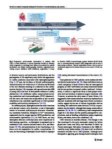
Methods DMD patients (n=236) were stratified into 4 groups (B-D, Figure 1) based on age, left ventricular (LV) ejection fraction (EF) and presence of myocardial fibrosis defined as positive myocardial delayed enhancement (MDE) compared to normal controls (group A, n=77). Dyssynchrony indices were calculated based on timing of CMR derived circumferential strain (ε cc ). The calculated indices included cross-correlation delay (XCD), uniformity of strain (US), regional vector of variance (RVV), time to maximum strain (TTMS) and standard deviation (SD) of TTMS. Abnormal XCD value was defined as > normal + 2SD. US, RVV, TTMS and SD were than derived for all patient population and patient with dyssynchrony defined as abnormal XCD. 1 CCHMC, Cincinnati, OH, USA Full list of author information is available at the end of the article
Figure 1
© 2011 Hor et al; licensee BioMed Central Ltd. This is an open access article distributed under the terms of the Creative Commons Attribution License (http://creativecommons.org/licenses/by/2.0), which permits unrestricted use, distribution, and reproduction in any medium, provided the original work is properly cited.
Hor et al. Journal of Cardiovascular Magnetic Resonance 2011, 13(Suppl 1):O17 http://jcmr-online.com/content/13/S1/O17
Page 2 of 2
Table 1 General Characteristic by Group Group Parameter
A (n=77)
B (n=90)
C (n=111)
D (n=19)
E (n
Data Loading...