Progressive Cervical Myelopathy as Presentation of Sarcoidosis
- PDF / 138,579 Bytes
- 2 Pages / 595.276 x 790.866 pts Page_size
- 64 Downloads / 336 Views
Progressive Cervical Myelopathy as Presentation of Sarcoidosis David Price, MD1, Richart Harper, MD2, and Mark C. Henderson, MD1 1
Department of Internal Medicine, University of California, Davis, Sacramento, CA, USA; 2Division of Pulmonary, Critical Care and Sleep Medicine, University of California, Davis, Sacramento, CA, USA.
J Gen Intern Med 28(6):855–6 DOI: 10.1007/s11606-012-2315-y © Society of General Internal Medicine 2013
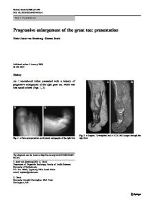
Figure 1. Cervical spine magnetic resonance imaging (MRI) of a 44-year-old African-American woman. Panel a: MRI with gadolinium contrast 3 weeks prior to admission. Panel b: MRI with gadolinium contrast after 8 weeks of corticosteroid therapy.
African-American woman was admitA ted44-year-old with four months of intermittent “shock-like” sensations in her chest radiating to both arms; she developed left arm weakness just prior to admission. A cervical spine magnetic resonance imaging (MRI) demonstrated intramedullary enhancement from C1 to T1 (Fig. 1 Panel a). Neurologic examination showed 4/5 strength of left wrist flexors and extensors, decreased bilateral light touch and pinprick sensation from C5 to T5, and hyperalgesia bilaterally from T1 to T3. Chest x-ray showed hilar adenopathy. Serum angiotensin converting enzyme level was 191 U/L (normal 9–67). Lumbar puncture was remarkable for a protein of 54 mg/dl (normal 15–45) and glucose of 30 mg/dl (normal 45–80;
serum glucose 106). Transbronchial biopsy showed noncaseating granulomas. Sarcoidosis is an idiopathic inflammatory disease characterized by non-caseating granulomas affecting any organ. Neurologic involvement occurs in 5–17 % of sarcoidosis patients, with 0.43 % having spinal cord involvement.1 The most common cerebrospinal fluid findings are elevated protein and hypoglycorrhachia, presumably a manifestation of chronic meningeal inflammation.2 Owing to the invasive nature of spinal cord biopsy, neurosarcoidosis is often diagnosed with extraneural tissue and the appropriate clinical syndrome.3,4 Treatment generally includes immunosuppressive agents. This patient’s neurological deficits and radiographic abnormalities improved with corticosteroids (Fig. 1 Panel b).
Received July 23, 2012 Revised October 18, 2012 Accepted December 12, 2012 Published online January 31, 2013
855
856
Price et al.: Progressive Cervical Myelopathy as Presentation of Sarcoidosis
Conflicts of Interest: David Price declares that he does not have a conflict of interest. Richart Harper receives grants from NIH R01 HL085311 (2008-13), NIH R01 HL096373 (2011-15), and NIH R01 HL105573 (2012-16). Mark Henderson receives grants from HRSA Primary Care Residency training D58HP05139 (2005-11). He also receives royalties from McGrawHill Medical Publishing, Book Royalties for The Patient History: an evidenced-based approach to differential diagnosis.
Corresponding Author: Mark C. Henderson, MD; Department of Internal Medicine, University of California, Davis, 4150 V Street, Suite 3100, Sacramento, CA 95817, USA (e-mail: [email protected]).
JGI
Data Loading...