Pulmonary thromboembolism with multiple right heart mural thrombus in a patient with COVID-19
- PDF / 969,303 Bytes
- 2 Pages / 595.276 x 790.866 pts Page_size
- 63 Downloads / 305 Views
CASE IMAGE IN CARDIOVASCULAR ULTRASOUND
Pulmonary thromboembolism with multiple right heart mural thrombus in a patient with COVID‑19 Houman Dehghan1 · Azam Soleimani1,2 Received: 24 August 2020 / Revised: 12 October 2020 / Accepted: 14 October 2020 © Japanese Society of Echocardiography 2020
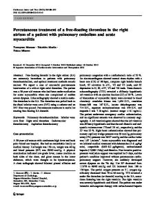
Hypercoagulation state during COVID-19 disease has been confirmed recently. Herein we present a 40-year-old hypertensive otherwise healthy male who was admitted with complaints of myalgia, low grade fever, dry cough, leg swelling, and exacerbating dyspnea in the last two weeks with history of recent recovery from COVID-19 in his wife. At presentation, he was afebrile with stable vital signs (blood pressure: 114/81 mmHg, respiratory rate: 16/minute, heart rate: 75 bpm) despite hypoxemia ( SPaO2 of 74% on room air, 94% on nasal O2 therapy). Coarse crackles in the lung fields, symmetric pitting leg edema and elevated jugular venous pressure were noticed in clinical exam with low voltage QRS, extreme right axis deviation and negative T waves in the precordial leads in electrocardiogram (Fig. 1a). Chest CT scan demonstrated pericardial and pleural effusions, filling defects in right atrium (RA) and right ventricle (RV) cavities, peripheral based ground glass opacities (compatible with COVID involvement), wedge-shaped pulmonary infarct and pulmonary arterial branch thrombosis (Fig. 1b, supplementary video1). No deep vein thrombosis was found. Transthoracic and transesophageal echocardiography showed preserved left ventricular systolic function, flattened interventricular septum, moderate right atrium (RA) and right ventricular (RV) enlargement and dysfunction, plethoric inferior vena cava with two large mural thrombi
Electronic supplementary material The online version of this article (https://doi.org/10.1007/s12574-020-00500-x) contains supplementary material, which is available to authorized users.
(4 cm × 2.5 cm) in RA appendage and RV apex (Fig. 1c, d, supplementary videos 2, 3). In laboratory data, positive oropharyngeal swab for severe acute respiratory syndrome corona virus 2 (SARS-COV2), lymphopenia (782 per µL) without leukocytosis (4150 per µL), elevated d-dimer (4510 ng/ml, normal
Data Loading...