Quantitative characterization of the three-dimensional microstructure of polycrystalline Al-Sn using X-ray microtomograp
- PDF / 599,214 Bytes
- 9 Pages / 607.68 x 788.88 pts Page_size
- 110 Downloads / 301 Views
5/25/04
11:55
Page 1953
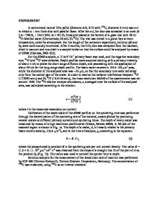
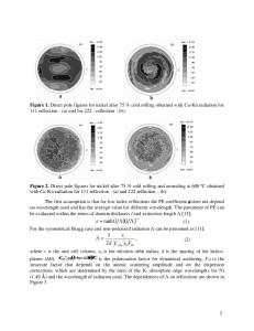
Quantitative Characterization of the Three-Dimensional Microstructure of Polycrystalline Al-Sn using X-Ray Microtomography K.M. DÖBRICH, C. RAU, and C.E. KRILL III Characterizing the three-dimensional topology of grain-boundary networks in polycrystalline materials is a crucial step in the modeling of properties that depend on the sample microstructure. Using absorptioncontrast X-ray microtomography, we have carried out a large-scale microstructural characterization of polycrystalline Al doped with 2 at. pct Sn, which is immiscible in Al in the solid state. The segregation of Sn to the grain boundaries imparts a strong contrast in X-ray attenuation that can be reconstructed tomographically; however, the nonuniformity of the segregation process presents a formidable challenge to the automated segmentation of the reconstructions. By employing an iterative grain-finding algorithm followed by a novel grain-boundary-network optimization routine (based on a phase-field simulation of grain growth), we were able to extract reliable values for the size and topology of nearly 5000 individual grains in polycrystalline Al-Sn. The distributions and averages of these data deviate significantly from the corresponding microstructural parameters of the three-dimensional (3-D) Poisson–Voronoi tessellation often used to model polycrystalline samples. Much better agreement was observed with microstructures generated by the computer simulation of three-dimensional grain growth.
I. INTRODUCTION
THE success of materials science as a scientific discipline rests to a great extent on the recognition that the properties of a material are determined largely by its microstructure. Essential to the study of structure-property relationships is the accurate and comprehensive characterization of microstructure[1]—an undertaking that has traditionally been carried out by optical or scanning electron microscopy applied to two-dimensional (2-D) sections of three-dimensional (3-D) samples or by transmission electron microscopy of very thin sections cut from bulk material. Although a great deal of information can be gleaned from such planar samples, there are a number of microstructural features that can be ascertained adequately only via a truly 3-D characterization procedure.[2,3] Examples of such features include the sizes, shapes, and spatial and orientation distributions of microstructural elements such as grains, precipitates, secondary phases, porosity, cracks, and grain boundaries.[4–7] Moreover, relationships between these elements, such as correlations and interconnectivities,[8,9] are inherently 3-D in nature and, therefore, difficult or even impossible to quantify on the basis of 2-D sections. In the past, the three-dimensionality of materials microstructures has been investigated primarily by the technique of serial sectioning,[2,10–14] which, owing to its tedious nature, has been performed rather infrequently.[2] Recent advances K.M. DÖBRICH, Doctoral Student, is with the Institut für Experimentalphysik, Freie
Data Loading...