Radial head fractures in young, active patients
- PDF / 1,141,661 Bytes
- 8 Pages / 595 x 792 pts Page_size
- 4 Downloads / 351 Views
Übersicht Obere Extremität https://doi.org/10.1007/s11678-020-00605-w Received: 12 August 2020 Accepted: 15 September 2020 © The Author(s) 2020
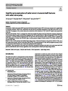
A fully functional upper extremity depends on the stability of the elbow joint. Fractures can compromise this stability and radial head fractures account for 30% of elbow fractures [1]. The incidence of radial head fractures has a bimodal pattern, with a peak at 37 years for men and 52 forwomen(. Fig. 1; [1–3]). The mechanism of injury is most commonly a fall onto an outstretched hand; however, any mechanism applying an axial force along the longaxisofthe radius canbe causative. Women tend to sustain lower-energy injuries, whereas males more often sustain higher-energy injuries [2]. Disruption of the important ligamentous stabilizers of the elbow joint are also common with radial head fractures.
A fully functional upper »extremity depends on the stability of the elbow joint Although extensively studied, treatment of radial head fractures remains controversial particularly in younger patients with more severe injuries [4]. Pain, stiffness, and limitations in range of motion (ROM) are associated with both operative and nonoperative treatment. As a result, these injuries can lead to significant disability in young, active patients if not managed appropriately.
Christopher G. Larsen1 · Michael J. Fitzgerald1 · Andrew S. Greenberg1,2 1 2
Department of Orthopaedic Surgery, Northwell Health, New Hyde Park, USA Orthopaedic Associates of Manhasset, Manhasset, USA
Radial head fractures in young, active patients is a secondary restraint to valgus forces on the elbow and provides longitudinal stability, preventing proximal migration of the radius. The concave radial head articulates with the convex capitellum and the medial rim of the radial head articulates with the lesser sigmoid notch of the ulna. Articular cartilage covers the radial head in a 280-degree arc with the remaining 80 degrees representing the nonarticular posterolateral side. This nonarticular “safe zone” can be identified as the region between two longitudinal lines drawn from the radial styloid and Lister’s tubercle [1, 3]. When using plate fixation, the plate should be placed in this safe zone to prevent impingement on the ulna during forearm pronation and supination. Given that ligamentous injury is common with radial head fractures, understanding the ligaments of the elbow and their contributions to stability is necessary for successful outcomes. On the lateral side of the elbow the lateral ulnar collateral ligament (LUCL) is the primary restraint to varus and external rotation forces and prevents posterolateral rotatory instability. Similarly, on the medial side the anterior bundle of the medial collateral ligament (MCL) is the primary restraint to valgus forces on the elbow, the posterior bundle, and the transverse bundle [1, 5].
Anatomy The elbow joint is made up of three articulations: ulnohumeral, radiocapitellar, and proximal radioulnar. Overall, 60% of the load transferred across the elbow joint is transmitt
Data Loading...