Relationship between mucoid hypertrophy of the anterior cruciate ligament (ACL) and morphologic change of the intercondy
- PDF / 231,286 Bytes
- 6 Pages / 595.276 x 790.866 pts Page_size
- 56 Downloads / 284 Views
SCIENTIFIC ARTICLE
Relationship between mucoid hypertrophy of the anterior cruciate ligament (ACL) and morphologic change of the intercondylar notch: MRI and arthroscopy correlation Ji Hyeon Cha & Sang Hoon Lee & Myung Jin Shin & Byeong Kyoo Choi & Sung Il Bin
Received: 12 November 2007 / Revised: 23 January 2008 / Accepted: 12 May 2008 / Published online: 16 July 2008 # ISS 2008
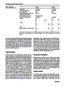
Abstract Objective The purpose of this study was to evaluate the relationship between mucoid hypertrophy of the anterior cruciate ligament (ACL) and morphologic change of the intercondylar notch. Materials and methods We retrospectively reviewed the 105 patients with knee magnetic resonance imaging (MRI) with or without knee arthroscopy [group 1: patients with arthroscopic notchplasty (N=47), group 2: knee arthroscopy demonstrating intact ACL (N=33), and group 3: patients with normal knee MRI but no arthroscopy (N=25)]. Groups 2 and 3 served as an arthroscopic and MR control group, respectively. Two musculoskeletal radiologists reviewed all MR examinations. The intercondylar notch width, notch index (width of intercondylar notch/width of femoral condyle), transverse notch angle (TNA), sagittal notch angle (SNA), and notch area were recorded on axial and sagittal MR images at the midpoint of Blumensaat’s line which was identified on sagittal images. The diameter of the ACL was recorded on coronal MR images at the posterior end of Blumensaat’s line. J. H. Cha : S. H. Lee : M. J. Shin : B. K. Choi Department of Radiology and Research Institute of Radiology, Asan Medical Center, University of Ulsan College of Medicine, Seoul, South Korea S. I. Bin Department of Orthopedic Surgery, Asan Medical Center, University of Ulsan College of Medicine, Seoul, South Korea S. H. Lee (*) Department of Radiology, Asan Medical Center, University of Ulsan College of Medicine, 388-1, Pungnap2-dong, Songpa-gu, Seoul 138-736, South Korea e-mail: [email protected]
Results The mean values of the intercondylar notch width, notch index, TNA, SNA, notch area, and ACL diameter for the three groups were 16.0 mm/0.2/50.3°/36.5°/249.0 mm2/ 7.7 mm (group 1); 19.3 mm/0.3/52.9°/40.2°/323.4 mm2/ 4.8 mm (group 2); and 20.3 mm/0.3/51.4°/39.1°/ 350.8 mm2/4.5 mm (group 3). The intercondylar notch width, notch index, SNA, and notch area were smaller, and ACL diameter was thicker in group 1 compared with the other groups (p
Data Loading...