Revascularization to the bone tunnel wall after anterior cruciate ligament reconstruction may relate to the distance fro
- PDF / 1,081,587 Bytes
- 7 Pages / 595.276 x 790.866 pts Page_size
- 54 Downloads / 277 Views
(2020) 32:53
Knee Surgery & Related Research
RESEARCH ARTICLE
Open Access
Revascularization to the bone tunnel wall after anterior cruciate ligament reconstruction may relate to the distance from the vessels Yuji Arai1, Kunio Hara2, Hiroaki Inoue3* , Hitoshi Kanamura3, Shuji Nakagawa1, Satoru Atsumi2 and Yasuo Mikami4
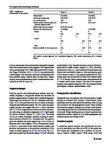
Abstract Purpose: We use magnetic resonance angiography to evaluate the difference of vascular ingrowth to the bone tunnel on the anterior and posterior walls quantitatively after anterior cruciate ligament reconstruction. Materials and methods: One hundred patients underwent anterior cruciate ligament reconstruction with multistranded semitendinosus tendons. They were retrospectively divided into those who underwent magnetic resonance angiography 2, 3, 4 to 6, and ≥ 7 months after surgery. The mean signal-to-noise ratios of the bone tunnel walls in the femur and tibia from the digital data were measured and compared for the anterior and posterior walls. Results: The signal-to-noise ratio of the posterior wall of the femoral bone tunnel was significantly higher than that of the anterior wall in each group. On the tibial side, the signal-to-noise ratio of the anterior wall was significantly higher than that of the posterior wall at ≥4 months after surgery. Conclusions: This study showed that the blood flow after anterior cruciate ligament reconstruction to the femoral bone tunnel is maintained from the posterior wall, and is maintained to the tibial side from the anterior wall 4 months postoperatively. Revascularization to the bone tunnel wall after anterior cruciate ligament reconstruction may relate to the distance from the vessels. Keywords: Anterior cruciate ligament, Bone tunnel wall, Magnetic resonance angiography, Revascularization
Introduction The anterior cruciate ligament (ACL) is a support mechanism responsible for stabilization of the knee joint. Recovery of ACL function after injury is needed because decline of knee joint function occurs with ACL injury and has a significant influence on both daily and sports activities. As surgical therapy, ACL reconstruction is * Correspondence: [email protected] 3 Department of Orthopaedics, Graduate School of Medical Science, Kyoto Prefectural University of Medicine, 465, Kajiicho, Kawaramachi-Hirokoji, Kamigyo-Ku, Kyoto, Kyoto 602-8566, Japan Full list of author information is available at the end of the article
widely performed, in which the graft is passed through bone tunnels made in the tibia and femur. For the tendon graft material, the hamstring, bone-patellar tendon, iliotibial tract, quadriceps femoris tendon, and allograft tissue are used [1–4]. A hamstring autograft is one of the most frequently used grafts and has shown favorable clinical results [4–9]. However, the ability to engage in sports activities may require a long time to return [10]. Remodeling processes, such as remodeling of the tendon graft and consolidation of the bone-tendon junction, are important as biological factors that determine the treatment outcome of ACL
Data Loading...