Right aortic arch with an aberrant left subclavian artery and aortic coarctation including a descending aortic aneurysm
- PDF / 1,024,089 Bytes
- 4 Pages / 595.276 x 790.866 pts Page_size
- 90 Downloads / 325 Views
(2019) 14:65
CASE REPORT
Open Access
Right aortic arch with an aberrant left subclavian artery and aortic coarctation including a descending aortic aneurysm Frantisek Sabol1, Peter Candik2*, Adrian Kolesar1 and Tomas Toporcer1
Abstract Backround: The right aortic arch and aortic coarctation are rare congenital anomalies with the incidence of 0.1% and 0.03–0.04%. We present a case report of a 51-year-old woman with the right aortic arch with aberrant left subclavian artery and coarctation of the aorta with post-stenotic aneurysm. Case presentation: Resection of the coarctation and aneurysm with replacement by tubular prosthesis was performed on partial cardiopulmonary bypass via femoral vessels. Conclusion: Partial cardiopulmonary bypass is an applicable method for ensuring the perfusion of the distal part of the body and an aberrant left subclavian artery is not a contraindication of this technique. Keywords: Aortic coarctation, Partial cardiopulmonary bypass, Right aortic arch
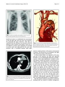
Introduction Asymptomatic congenital anomalies with an incidence of 0.5–3% include left aortic arch with an aberrant right subclavian artery with or without diverticulum of Kommerell, double site ligamentum arteriosum or ductus arteriosus and left circumflex aorta [1, 2]. Right aortic arch (RAA) usually comprises an aberrant left subclavian artery most commonly with retroesophageal diverticulum. It is often associated with left sided ductus arteriosus and vascular ring [1]. Symptomatic aortic arch anomalies contain hypoplasia, coarctation (CoA) and pseudocoarctation of the aorta [1]. Aortic arch anomalies can occur in an isolation and with other congenital anomalies as well. In 1948, Edwards explained the various congenital anomalies by system of embryonic double aortic arch and site of interruption or atresia of the embryonic arch system [2]. RAA occurs in 0.1% of adults [2]. Regression of 4th left aortic arch can occurs in different part of the arch with different finally anatomy composition (Fig. 2a and b). On the other hand, CoA presents 5–8% of all * Correspondence: [email protected] 2 Clinic of Anaesthesiology and Intensive Medicine, Medical Faculty of P.J.Safarik University and Eastern Slovak Institute for Cardiovascular Diseases Ltd, Ondavska 8, 040 01 Kosice, Slovakia Full list of author information is available at the end of the article
congenital heart defects with estimated incidence of 0.3–0.4 per 1000. It can by solitary in 82% of humans or associated with other congenital malformations and symptoms. Late aneurysmal formation in the distal CoA is one of the complications due to untreated CoA with high risk of aortic rupture and death [3].
Case report A 51-year-old woman with history of RAA in surveillance attended a cardiologist due to dyspnea and palpitation. The patient underwent a computer tomography (CT) evaluation which confirmed RAA with a common origin of both carotid arteries, a separate origin of the right subclavian artery, coarctation of the aorta with the diameter of 12 mm, an aortic aneurysm below the coarcta
Data Loading...