Scoliosis in spinal muscular atrophy: is the preoperative magnetic resonance imaging necessary?
- PDF / 505,099 Bytes
- 3 Pages / 595.276 x 790.866 pts Page_size
- 70 Downloads / 304 Views
CASE SERIES
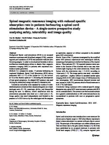
Scoliosis in spinal muscular atrophy: is the preoperative magnetic resonance imaging necessary? Nestor Ricardo Davies1 · Eduardo Galaretto1 · Lucas Piantoni1 · Rodrigo Germán Remondino1 · Ida Alejandra Francheri Wilson1 · María Soledad Monges1 · Sofía Frank1 · Ernesto Salomón Bersusky1 · Carlos Alberto Tello1 · Mariano Augusto Noel1 Received: 12 March 2020 / Accepted: 4 May 2020 © Scoliosis Research Society 2020
Abstract Purpose To determine the prevalence of intraspinal alterations in scoliosis due to Spinal Muscular Atrophy (SMA). Methods Cross-sectional, observational, descriptive study. Fifty-six patients with SMA diagnosis required surgical treatment due to scoliosis. Inclusion criteria: scoliosis/kyphoscoliosis > 50 degrees in the coronal plane, clinical characteristics of Spinal Muscular Atrophy, accurate diagnosis by means of molecular or genetic study. Prior to the spinal surgery, and to find related intraspinal alterations, MRI of the spine and posterior cranial fossa was performed. Results Forty females, 16 males, mean age 11 years (range 6–14 years). 94% of the patients had Spinal Muscular Atrophy type 2. The mean angle value was 81 degrees (range 53–122 degrees) in the coronal plane and 62 degrees (range 35–80 degrees) in the sagittal plane. The prevalence of intraspinal alterations was 1.78%. One patient with cervical hydromyelia and no neurological surgical procedure prior to the spinal deformity surgery was reported. Conclusions In the context of preoperative planning and strategy of patients with scoliosis due to Spinal Muscular Atrophy, MRI may have not to be requested. Keywords Scoliosis · Spinal muscular atrophy · Magnetic resonance imaging
Introduction Spinal muscular atrophy (SMA) is a genetic disorder of autosomal recessive inheritance in 95% of the cases. The disorder is characterized by the loss of motor neurons of the anterior horns of the spinal cord causing muscle atrophy leading to proximal muscle and trunk weakness. The estimated incidence is 1 in 11,000 live births. The natural history of SMA is complex, variable, and multifactorial. Based on the clinical features and age at diagnosis, the disease has been classified into different types and subtypes [1]. Type 0: hypotonic neonates with severe respiratory involvement and a life expectancy of less than 6 months; type 1 or Werdnig–Hoffman disease: infants younger than 6 months of life who cannot sit without support; type 2: children younger than 18 months who, although they are able to sit, cannot * Nestor Ricardo Davies [email protected] 1
stand or walk independently; type 3 or Kugelberg–Welander disease: patients are generally older than 18 months and are able to walk without support at some point in their life; type 4: adult patients with the least severe form of the disease who walk independently [1, 2]. The incidence of scoliosis, one of the different orthopedic disorders associated with the disease, is between 50 and 70%. SMA-associated scoliosis is progressive in nature and the majority of patient
Data Loading...