Severity of mitral valve prolapse is associated with basal left ventricular hypertrophy: a cardiac magnetic resonance st
- PDF / 164,174 Bytes
- 2 Pages / 595.28 x 793.7 pts Page_size
- 110 Downloads / 309 Views
POSTER PRESENTATION
Open Access
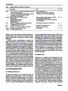
Severity of mitral valve prolapse is associated with basal left ventricular hypertrophy: a cardiac magnetic resonance study Mohammad I Zia1,2*, Valentina Valenti1, Caroline Cherston1, Maressa C Criscito1, Seth Uretsky1,3, Steven D Wolff1 From 15th Annual SCMR Scientific Sessions Orlando, FL, USA. 2-5 February 2012 Summary Mitral valve prolapse (MVP) is associated with concentric basal hypertrophy of the left ventricle. We found a strong correlation between the excursion of the mitral valve annulus and the degree of relative hypertrophy suggesting the possibility that locally increased myocardial function may be responsible for the hypertrophy. Background Recently, basal left ventricular hypertrophy has been suggested as a new form of hypertrophic cardiomyopathy. However, we have noticed that substantial focal basal hypertrophy often occurs in patients with mitral valve prolapse (Figure 1). Our objective was to characterize the extent and distribution of focal basal left ventricular hypertrophy in patients with MVP and assess the correlation between the degree of focal hypertrophy and various myocardial structural parameters.
Results Fourty-three (70%) patients had posterior leaflet prolapse, 2 (3%) patients had anterior leaflet prolapse and 17 (27%) patients had bileaflet prolapse. There was a significantly increased ratio of basal to mid end-diastolic wall thickness in all segments of the left ventricle in MVP patients when compared to controls (Figure 2). The inferolateral (2.1 vs. 1.0, p
Data Loading...