Simultaneous TEM and Cathodoluminescence Imaging of Non Uniformity in In 0.1 Ga 0.9 N Quantum Wells
- PDF / 271,511 Bytes
- 6 Pages / 612 x 792 pts (letter) Page_size
- 37 Downloads / 294 Views
L11.12.1
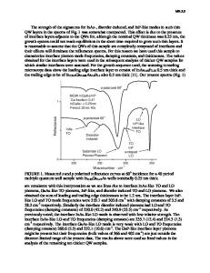
Simultaneous TEM and Cathodoluminescence Imaging of Non Uniformity in In0.1Ga0.9N Quantum Wells Nicholas M. Boyall1, Ken Durose1, and Ian M. Watson2 1 Department of Physics, University of Durham, South Road, Durham, DH1 3LE, UK. 2 Institute of Photonics, University of Strathclyde, Wolfson Centre, 106 Rottenrow, Glasgow, G4 ONW, UK. ABSTRACT Monochromatic cathodoluminescence (CL) imaging of metal-organic vapour phase epitaxy (MOVPE) grown In0.1Ga0.9N single quantum wells (QW) has been performed in a scanning transmission electron microscope (STEM). Spatially resolved fluctuations in the CL emission wavelength and intensity of the QW luminescence were recorded. The presence of regions with luminescent features asymmetrically distributed either side of the QW peak emission was inferred. These fluctuations may be attributed to, by for example, variations of ±0.01 in the In fraction of the In0.1Ga0.9N alloy, to changes of up to 0.6nm in the QW thickness. However these factors do not explain the gross fluctuations in QW emission intensity observed in TEM-CL on the scale of ~1µm. INTRODUCTION Light emitting and laser diodes based on InxGa(1-x)N quantum well (QW) structures have been available commercially for a number of years [1]. However the exact relationship between compositional distributions within the QWs and the efficiency and wavelength of light emission is not fully described. A number of authors describe an emission process based around the localisation of excitation to QW composition fluctuations [2-4]. As means of investigating non uniformity, emission from InxGa(1-x)N QW structures has been investigated by CL microscopy in the scanning electron microscope (SEM) by a number of authors [5-7]. More recently this work has been extended by measurement of CL in transmission electron microscopes with scanning attachments (STEM) [8, 9]. Measurement of CL in a STEM was first reported by Petroff et al [10] and has been reviewed elsewhere [11]. This method allows spectroscopic and microstructural information to be collected simultaneously. High spatial resolution can be achieved with this technique as the beam broadening in electron transparent TEM foils is small [12], and the effective carrier diffusion length is small due to the proximity of the foil surface. These advantages are traded off against the smaller signal available from a thin foil as a result of losses due to surface recombination and the limits of a small generation volume. In this work panchromatic and monochromatic STEM-CL imaging and spectroscopy were used to investigate the uniformity of emission from an InxGa(1-x)N /GaN single QW. EXPERIMENTAL DETAILS Specimens The single QW specimens were grown by metal-organic vapour phase epitaxy (MOVPE) on sapphire (0001) substrates. A GaN buffer layer approximately one micron thick was deposited
L11.12.2
on the substrate following deposition of a low temperature nucleation layer. The InxGa(1-x)N QWs were grown at 832ºC, with nominally 2.5nm thickness and were capped with 15nm of GaN. Further detail
Data Loading...