Structure of Highly Perfect Semiconductor Strained-Layer Superlattices: High-Resolution X-Ray Diffraction and Computer S
- PDF / 258,593 Bytes
- 5 Pages / 420.48 x 639 pts Page_size
- 1 Downloads / 342 Views
STRUCTURE OF HIGHLY PERFECT SEMICONDUCTOR STRAINED-LAYER SUPERLATTICES: HIGH-RESOLUTION X-RAY DIFFRACTION AND COMPUTER SIMULATION STUDIES
J. M. VANDENBERG AT&T Bell Laboratories, Murray Hill, New Jersey 07974.
ABSTRACT High-resolution x-ray diffraction (HRXRD) measurements of strained-layer superlattices (SLS's) have been carried out using a four-crystal monochromator. A wide asymmetric range of sharp higher-order x-ray satellite peaks is observed indicating well-defined periodic structures. Using a kinematical diffraction step model very good agreement between measured and simulated x-ray satellite patterns could be achieved. These results show that this x-ray method is a powerful tool to evaluate the crystal quality of SLS's.
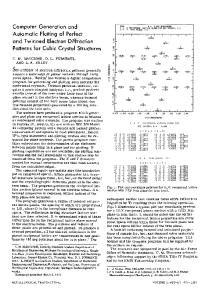
High-resolution x-ray diffraction (HRXRD) has been used to very precisely determine [1] the structural parameters of a number of strained-layer superlattices (SLS's) of various semiconductor materials. The high resolution was achieved with a four-crystal monochromator [2] and consists of an identical pair of crystals channel cut parallel to the (220) planes and in each block the x-rays go through two (220) reflections. This provides a highly parallel monochromatic CuKaj x-ray beam. This compact monochromator with the sample as the fifth crystal and an open end detector is arranged on a Huber 424/511 four-circle diffractometer. This x-ray method has been very successful in analyzing [1,3,4] SLS's grown by gas-source molecular beam epitaxy (GSMBE), which enables one to grow superlattices with a large degree of structural perfection, such as sharp interfaces, very thin layers and excellent control of composition [5]. The upper trace in Fig. la shows the 400 scan of a 20A InXGal_xAs/300AInP SLS with nominal composition x = .075, grown on (100) InP. The perpendicular lattice mismatch AaL/alnp can be obtained from the angular difference of the main (n = 0) superlattice (SL) and the substrate peak, and the period A from the positions of the satellite peaks. The presence of a large number of satellite peaks, up to order n = +10, indicates a well-defined periodic structure. The strong asymmetry in the satellites intensities, which shift to positive order number n, gives evidence of a strong negative strain as a result of the very low In concentration x as compared to x = 0.53 for a perfectly matched SL. This x-ray scan can be analyzed by using a kinematical step model [6,7] which assumes ideally sharp interfaces and calculates the diffracted amplitude F of the structural periodicity of the superlattice along the [100] growth direction. The variable input parameters of the step model are the number of molecular layers Nw and NB in the InGaAs well (W) and InP barrier (B), respectively, and their corresponding lattice spacings dw and dB, which are the distances between atomic layers along the 11001 direction and where one molecular layer =- 2 d. For alternating wells and barriers the diffracted amplitude F in the vicinity (h=4) of the (400) reflection is given by
Mat. Res. Soc. Symp. Proc. Vol. 145.
Data Loading...