Surgical anatomy in intersphincteric resection
Recently, surgical techniques for low rectal cancer have greatly progressed, thereby increasing the rate of sphincter preservation. As a result of the development and spread of surgical procedures including the total mesorectal excision (TME) [1] and inte
- PDF / 634,763 Bytes
- 7 Pages / 595 x 791 pts Page_size
- 39 Downloads / 306 Views
Introduction Recently, surgical techniques for low rectal cancer have greatly progressed, thereby increasing the rate of sphincter preservation. As a result of the development and spread of surgical procedures including the total mesorectal excision (TME) [1] and intersphincteric resection (ISR) [2], rectal surgeons more frequent have the opportunity to observe the anorectal junction and to mobilize the distal rectum to the anal canal. Knowledge about the anatomical structures in the lower pelvis is important for the abdominal and perineal parts of ISR. However, critically controversial descriptions of the fascial structures in the pelvis are common, except for the concept that the mesorectum is surrounded by visceral fascia or the fascia propria of the rectum [3]. Controversies exist with regard to which fascia covers the hypogastric nerve (HGN) and whether the rectosacral fascia is a real structure or a surgical artifact due to adhesion of the fasciae. Moreover, it is unclear whether the presacral fascia and Waldeyer’s fascia are the same fascia [4]. As Range and Woodburne [5] pointed out the possibility that pelvic fascial structures are easily developed during dissection and surgery, a comprehensive histological study using large sections covering wide areas around the mesorectum seems necessary. We performed histological studies to try and resolve the aforementioned controversies. It is important to thoroughly understand the morphology of the connective tissue structures around anal canal in order perform ISR and many other procedures. The rectourethralis muscles and the anococcygeal ligament are important structures at the anorectal junction for ISR. These structures are located between surgical planes by both the abdominal and peranal approach. The following descriptions will be useful for surgeons performing ISR and other procedures that affect the same region.
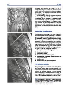
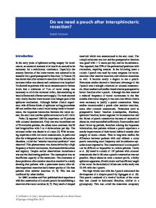
Fascial structures around the rectum (Figs. 1, 2) To avoid presacral venous bleeding, autonomic nerve injury and local recurrence, mobilization of the rectum is performed by anatomical dissection along the fascial planes [6, 7]. Therefore, a comprehensive understanding of the fascial structures around the rectum is critically important for rectal surgeons.
Fascia propria of the rectum The fascia propria of the rectum is a thin visceral fascia covering the rectum and mesorectum. The mesorectum is a distinct compartment that contains the superior rectal arteries and veins, mesorectal fat, lymphatic vessels and nodes. This fascia is also called the perirectal fascia, rectal fascia, and visceral fascia.
Denonvilliers’ fascia (Fig. 3) Denonvilliers’ fascia is clearly identifiable in males between the fascia propria of the rectum and the seminal vesicles or prostate. The rectovaginal septum in females corresponds to Denonvilliers’ fascia. The consistency of Denonvilliers’ fascia varies between individuals, from a fragile translucent layer to a tough leathery membrane [8]. The rectovaginal septum is less prominent in females than Denonvilliers’ fascia is
Data Loading...