Synchrotron X-ray Observations of a Monolayer Template for Mineralization
- PDF / 211,136 Bytes
- 6 Pages / 611.998 x 791.997 pts (letter) Page_size
- 91 Downloads / 262 Views
Synchrotron X-ray Observations of a Monolayer Template for Mineralization E. DiMasi,1∗ and L. B. Gower2 1 Dept. of Physics, Brookhaven National Laboratory, Upton NY 11973 2 Dept. of Materials Science and Engineering, University of Florida, Gainesville FL 32611 ∗ Corresponding author: [email protected]
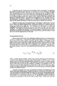
ABSTRACT Mineral nucleation at a Langmuir film interface has been studied by synchrotron x-ray scattering. Diluted calcium bicarbonate solutions were used as subphases for arachidic and stearic acid monolayers, compressed in a Langmuir trough. Self-assembly of the monolayer template is observed directly, and subsequent crystal growth monitored in-situ. INTRODUCTION A persistent question in biomineralization concerns the interactions between the organic molecules and the nucleating mineral. Since mineralization often occurs in proximity to an organic matrix having structural regularities that match the mineral lattice spacings, the organic material is assumed to act as a template for nucleation [1]. However, a number of recent experiments emphasize that mineralization can also proceed through amorphous presursors, which subsequently crystallize [2, 3]. In these systems, the importance of registry with a macromolecular template is called into question. One important model system that addresses this issue consists of a surfactant monolayer assembled at the air–water interface. These molecules are well known to form ordered, two-dimensional templates, typically with centered rectangular or hexagonal lattices [4]. With the right choice of surfactant, the intermolecular distances, headgroup charges, and so on can be systematically varied. The advantages of these tunable systems have been demonstrated in the study of calcium carbonate nucleation, where calcite or vaterite formation with specific crystal size distribution, habit, and orientation at the surface was observed in the presence of various surfactants [5]. Similar work has been pursued with other minerals [6, 7]. In the works cited above, crystal morphology was assessed by optical and electron microscopy. These observations were not sensitive to the microscopic arrangement of surfactant molecules and cations, nor could they directly measure crystal growth during its earliest stages. In fact, this has been a common limitation for other studies of biomineralizing systems: detailed measurements of lattice spacings of mineral and template during early growth times have not been available. Similarly, the potential for mineralization to proceed through metastable phases such as amorphous or hydrated precursors can not always be assessed. In this paper we describe synchrotron x-ray scattering studies of calcium carbonate nucleating from solution at fatty acid monolayers. Our aim is to give an overview of the structural information that can be directly obtained by this method. Possible routes to mineralization in this system are illustrated in Figure 1. First, it is expected that the
HH3.38.1
a
b
c
d
Figure 1. (a) Fatty acid monolayer assembled on an aqueous calcium bic
Data Loading...