Test yourself answer to question: Incidental left L5/S1 facet finding on MRI
- PDF / 916,747 Bytes
- 2 Pages / 595.276 x 790.866 pts Page_size
- 6 Downloads / 321 Views
TEST YOURSELF: ANSWER
Test yourself answer to question: Incidental left L5/S1 facet finding on MRI Ramanan Rajakulasingam 1
&
Daniel Lindsay 2 & Hanny Anwar 3 & Asif Saifuddin 1 & Thillainayagam Muthukumar 1
Received: 16 August 2020 / Revised: 27 September 2020 / Accepted: 30 September 2020 # ISS 2020
Diagnosis Primary aneurysmal bone cyst of the left S1 superior articular process.
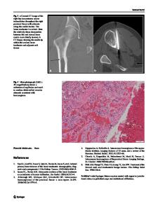
Discussion MRI (Fig. 1) shows a low T1- and heterogeneous T2weighted (T2W) signal intensity (SI) lesion arising from an expanded left S1 superior articular process. The L5/S1 facet joint was markedly irregular with multiple small cystic spaces evident on the T2W image, but no appreciable fluid-fluid levels. CT (Fig. 2) confirms a lytic lesion arising from the left S1 superior articular process with an egg-shell thin blown-out cortical appearance and no internal matrix mineralisation. The working differential diagnosis included osteoblastoma, fibrous dysplasia and less likely telangiectatic osteosarcoma.
Plain radiographs were unfortunately not performed. Although the lumbar region was asymptomatic, the rather unusual appearances warranted a CT biopsy with histology (Fig. 2) revealing blood-filled bony trabeculation with scattered osteoclast-like giant cells and foci of metaplastic ossification (*). Spindle cells embedded in a fibrous stroma lined the wall consistent with primary aneurysmal bone cyst (ABC) with no atypical mitoses. Most spinal ABC patients are less than 20 years old with a slight female predilection, the majority presenting with slowonset chronic back pain and a minority having underlying scoliosis. Some authors state a lumbar [1], while others report a cervical and thoracic predominance [2] for spinal ABC’s. In a case series of 14 lumbar spine ABC’s by Kleuver et al. [3], 9
The case presentation can be found at doi: 10.1007/s00256-020-03638-y * Ramanan Rajakulasingam [email protected] Daniel Lindsay [email protected] Hanny Anwar [email protected] Asif Saifuddin [email protected] Thillainayagam Muthukumar [email protected] 1
Department of Radiology, Royal National Orthopaedic Hospital, Brockley Hill, Stanmore, Middlesex HA7 4LP, UK
2
Department of Histopathology, Royal National Orthopaedic Hospital, Brockley Hill, Stanmore, Middlesex HA7 4LP, UK
3
Department of Orthopaedic surgery, Royal National Orthopaedic Hospital, Brockley Hill, Stanmore, Middlesex HA7 4LP, UK
Fig. 1 Axial T2W FSE MR image showing irregular expansion of the left S1 superior articular process, which contains multiple small cystic areas but no convincing fluid-fluid levels (arrow)
Skeletal Radiol
Fig. 2 Microphotographs a H&Ex10 and b H&Ex2. Histologically, a shows tumour composed of bland, relatively monomorphic spindle cells with a fibroblastic morphology arranged in a vague storiform architecture and embedded in a fibrous stroma. There are scattered osteoclast-like giant cells present and foci of metaplastic ossification (*). Importantly,
there is no significant cytological atypia and atypical mitoses.
Data Loading...