The diagnostic challenge of cardiomyopathies: When should we suspect cardiac amyloidosis?
- PDF / 6,313,427 Bytes
- 6 Pages / 593.972 x 792 pts Page_size
- 3 Downloads / 309 Views
Department of Cardiovascular Department of Department of
Nuclear Cardiology, Ramos Mejı´a Hospital, Buenos Aires, Argentina Institute of Buenos Aires, Buenos Aires, Argentina Cardiology, Ramos Mejı´a Hospital, Buenos Aires, Argentina Echocardiography, Ramos Mejı´a Hospital, Buenos Aires, Argentina
Received Sep 30, 2019; accepted Oct 1, 2019 doi:10.1007/s12350-019-01922-6
INTRODUCTION Cardiomyopathies constitute a heterogeneous group of diseases that produce different degrees of dysfunction. The etiological causes are multiple and are often combined in the same patient. The diagnosis of cardiac amyloidosis may be underestimated in patients with chronic coronary artery disease as both conditions share similar clinical manifestations. Cardiac amyloidosis is more common in elderly people in whom the prevalence of coronary artery disease is also high.1 The prevalence of cardiac amyloidosis with coronary artery disease is unknown. CASE SUMMARY This is a 74-year-old male patient with a history of hypertension. He had no history of coronary artery disease. He was hospitalized due to class II to III dyspnea associated with edema in both lower extremities. The initial diagnosis was heart failure with unknown etiology.
Reprint requests: Magali Gobbo, Department of Nuclear Cardiology, Ramos Mejı´a Hospital, General Urquiza 609, 1221 Buenos Aires, Argentina; [email protected] J Nucl Cardiol 1071-3581/$34.00 Copyright Ó 2019 American Society of Nuclear Cardiology.
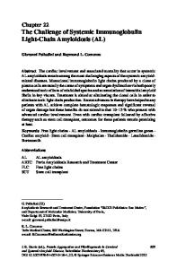
ECG showed sinus rhythm, low voltages in limb leads, septal inactivation, and frequent ventricular premature beats. The echocardiogram was suggested of infiltrative cardiomyopathy. However, it showed moderate LV dysfunction with wall motion abnormalities (Figure 1). The heart team was confused with one group suspected of advanced cardiac amyloidosis and the other group suspected of CAD. To establish the differential diagnosis between both etiologies, the patient underwent a 99mTc—MIBI SPECT (Figure 2). 72 hours later, the patient underwent 99mTc- HMDP cardiac scintigraphy that was positive (Figure 3)2,3. Both scintigraphies were compared (Figure 4). Finally, the patient underwent coronary angiography which revealed severe stenosis in the LM and 3VD (Figure 5). We report the case of a patient with transthyretin cardiac amyloidosis associated with CAD, with clinical and imaging features suggestive of both entities, reflecting the difficulty and the wide range of diagnostic tests
Gobbo et al The diagnostic challenge of cardiomyopathies: when should we suspect cardiac amyloidosis?
Journal of Nuclear CardiologyÒ
Figure 1. Echocardiogram on admission. A: Four-chamber view shows dilation of both atria, with thickened interatrial septum (blue arrow), normal LV diameters with severe increase in LV mass and sparkling appearance of the myocardium, and increased septal wall thickness with granular pattern (green arrow). B Four-chamber view shows moderate LV dysfunction with wall motion abnormalities with akinesia of the lateral segments. C Four-chamber view shows no
Data Loading...