The Integument and Moult
The integument covered by the cuticle has been studied in recent years in great detail thanks to the introduction of the scanning electron microscope (SEM). This has enabled the observation of the fine microstructure, and the various sensory structures as
- PDF / 1,520,939 Bytes
- 9 Pages / 439.37 x 666.142 pts Page_size
- 14 Downloads / 281 Views
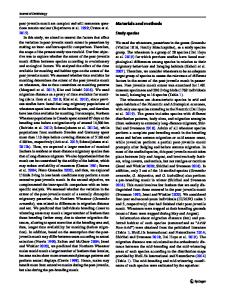
2.1 Structure and Function The integument covered by the cuticle has been studied in recent years in great detail thanks to the introduction of the scanning electron microscope (SEM). This has enabled the observation of the fine microstructure, and the various sensory structures associated with integumental covering (Figs. 2.1-2.14). Larger tubercles (Figs. 2.1-2.6) could be seen, and various functions assigned to them such as protection and water conservation (Schmalfuss 1975, 1977). There is some relationship between morphological and environmental or behavioural adaptations of isopods (Schmalfuss 1984). Various plaques and pits (Figs. 2.7-2.10) as well as setae and sensillae (Figs. 2.11, 2.13) have been described (Holdich and Lincoln 1974). All terrestrial isopods examined have numerous tricorn-shaped sensillae on their tergites (Figs. 2.12, 2.13). These sensillae are dispersed among plaques of various forms and pits in between. The tricorns are scale-like structures, and the plaques partly overlap, similar to a tile roof. The pits are semicircular depressions. The tricorns seem to be innervated and could be hygroreceptors (Price and Holdich 1980a). The setae observed at the margins of the tergite plates could be proprioceptors (Fig. 2.14). Similar observations were made on several isopod species by Schmalfuss (1975, 1977, 1978a), Powell and Holcrow (1982) and Holdich (1984). The integument consists of the epidermis, a lamellated procuticle (or endocuticule) and a two-layered epicuticle (Price and Holdich 1980a). Recently, Compere (1991) has described the fine structure of Oniscus asellus cuticle. The epicuticle is composed of five layers: (1) a cement layer with numerous dermal canal ducts; (2) a surface coat; (3) four laminated cuticulin layers; (4) a wax layer; and (5) the inner epicuticle. The outer layer of the epicuticule contains lipids in some isopod species (Hemilepistus reaumuri; see Hadley and Warburg 1986; Chap. 6). In Porcellionides pruinosus the epicuticule is covered by numerous spherical particles or balls of various diameter (Figs. 2.6-2.9). One possible function of these balls could be to reduce transpiration (Hadley and Hendricks 1985). All these cuticular structures contain Ca 2 +. This calcium carbon-
M. R. Warburg, Evolutionary Biology of Land Isopods © Springer-Verlag Berlin Heidelberg 1993
Structure and Function
2.1
5
2.2
2.3
2.4
Fig. 2.1. Head region of Bathytropa wahrmani showing tubercles ( x 75) Fig. 2.2. A single tubercle, enlarged ( x 350) Fig. 2.3. Tubercles of Porcellio barroisi ( x 200) Fig. 2.4. A single tubercle as in Fig. 2.3, enlarged ( x 500)
6
The Integument and Moult
ate in the integument of Oniscus asellus is amorphous (Wood and Russell 1987). In the integument, under the epidermal cell layer, lies the chromatophore layer, which contains spherical pigment granules that change during the isopod's development (Negishi and Hasegawa 1991).
2.1.1 The Tegumental Glands Tegumental glands were observed in terrestrial isopods about 100 years ago ("Webers glands", see
Data Loading...