The Use of Image Analysis in Long-Term Nondestructive Corrosion Monitoring of Nuclear Alloys
- PDF / 2,980,360 Bytes
- 12 Pages / 414.72 x 648 pts Page_size
- 120 Downloads / 268 Views
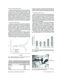
THE USE OF IMAGE ANALYSIS IN LONG-TERM NONDESTRUCTIVE CORROSION MONITORING OF NUCLEAR ALLOYS P.J.B. SCOTT *, S. SANDLOEHKEN **AND GERARD GUERIN *** CARIAD Consultants, 397 Parkview Avenue, North York, Ontario, Canada M2N 3Z7 Ontario Hydro Research Division, 800 Kipling Avenue, Toronto, Ontario, Canada M8Z 5S4 ".. Centre de Caracterisation Microscopique des Materiaux, Ecole Polytechnique, Universite de Montreal, Quebec, Canada
INTRODUCTION Direct examination of materials suspected of damage has been found to be the most accurate and reliable method of examination. It is labour intensive, however, and relies heavily on the experience and judgement of the operator. Furthermore, it is not always feasible to remove tubes and cut them into small fragments for such examination, particularly in nuclear applications. Nuclear waste packages for both high and low level waste will be made of materials that are not particularly vulnerable to pitting under anticipated conditions. Consequently, it will be necessary to make observations on specimens that contain relatively few significant pits scattered over relatively large areas, and it may be necessary to examine test specimens with curved surfaces. Cost considerations suggest that test techniques will be needed that combine image analysis with some sort of replication technique, so as to permit the collection of pit-distribution data. It is conceivable that some specimens will be replicated and then further corroded. Obviously, the process of replication will disturb the corrosion chemistry, but such a technique could permit the mapping of large areas in an impractical way, using existing vibrating-electrode techniques. Replication of areas of interest using acetate peels provides a rapid, inexpensive, and non-destructive alternative to removal of the sample. It also results in an easily stored permanent record of the damaged area for later comparison. It is important, however, that the method not give misleading results on the extent of damage. Image analysis, combined with optical and scanning electron microscopy (SEM), was assessed as a potential method for quantitative analysis of both surface cover by microorganisms and corrosion of the metal surface by these organisms. To test the reliability of the results using replicating techniques in combination with the image analyzer, a "standard" sample was examined by three different methods: direct analysis of the sample with image analysis, image analysis of an untreated peel, and image analysis of a peel from a sample treated with penetrant. This last method was divided further into analysis of various candidate penetrants. Direct analysis was used as a reference by which to judge the other methods. The important consideration was to differentiate surface roughness of the six alloys (which differs among the various alloys) from pit initiation. The threshold of sensitivity must be determined to include the smallest real pits possible, while excluding natural surface irregularities not caused by corrosion. Techniques were asse
Data Loading...