Transient left ventricular systolic dysfunction mimicking myocardial infarction after pericardiocentesis
- PDF / 146,582 Bytes
- 3 Pages / 595.276 x 790.866 pts Page_size
- 38 Downloads / 380 Views
CASE REPORT
Transient left ventricular systolic dysfunction mimicking myocardial infarction after pericardiocentesis R. W. J. H. Weijers & J. C. Post
Published online: 8 May 2013 # The Author(s) 2013. This article is published with open access at Springerlink.com
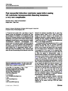
Case A 69-year-old woman was admitted to our hospital because of weight loss and progressively worsening dyspnoea. Her previous medical history was unremarkable. She was admitted to the internal medicine department. A cardiologist was consulted because of atrial fibrillation with rapid ventricular response and an enlarged cardiac silhouette on her chest Xray. On physical examination her blood pressure was 120/70 mmHg with a heart rate of 120 beats/min. She had engorged jugular veins and an enlarged liver without peripheral oedema. Her ECG showed atrial fibrillation and low voltage. No Q waves were noted. An echocardiogram showed a large pericardial effusion of more than 30 mm and a swinging heart (Fig. 1). The left ventricle was small and hyperdynamic without wall motion abnormalities. There was no collapse of the right ventricle. The inferior vena cava was dilated without inspirational collapse. Because of imminent cardiac tamponade a pericardiocentesis was performed with the immediate evacuation of 800 cc of haemorrhagic pericardial fluid. Six hours after this procedure control echocardiography was performed which showed only a small amount of pericardial fluid, but now a poor left ventricular function with general hypokinesia and anterior and septal akinesia. T-wave inversion and Q waves were seen on the ECG in the anterolateral leads (Fig. 2) but the cardiac markers were not elevated. She was treated with ACE inhibitors, diuretics and low-dose beta blockade. Analysis of the pericardial fluid showed malignant cells and on a chest CT scan a mass was detected. Subsequently, she was diagnosed with small cell lung carcinoma (SCLC) stage IV R. W. J. H. Weijers (*) : J. C. Post Department of cardiology, Catharina Hospital Eindhoven, PO Box 1350, 5602 ZA Eindhoven, the Netherlands e-mail: [email protected]
with metastasis to the pelvis, pericardium and brain and treated with radiation therapy. She was discharged and returned several months later with complaints of fatigue and progressive dyspnoea. Echocardiography again showed a large pericardial effusion; the left ventricle now had a normal systolic function without wall motion abnormalities. The ECG now showed normal R progression in the anterior leads with disappearance of Q waves (Fig. 3). She was again treated with pericardiocentesis and was discharged the following day. After this episode she died at home as a consequence of her progressive lung carcinoma.
Discussion Pericardiocentesis for cardiac tamponade is a commonly performed procedure which may lead to complications such as perforation of the (right) ventricle, laceration of a coronary artery, pneumothorax and ultimately death. Transient left ventricular dysfunction has been described by various authors [1–7]. Case reports describe patients
Data Loading...