Ventricular tachycardia originating from the crux of the heart treated with ablation within the cardiac venous system in
- PDF / 514,616 Bytes
- 2 Pages / 595.276 x 790.866 pts Page_size
- 75 Downloads / 286 Views
CASE REPORTS
Ventricular tachycardia originating from the crux of the heart treated with ablation within the cardiac venous system in a 12-year-old patient Paola Ferrari 1 & Giovanni Malanchini 1
&
Cristina Leidi 1 & Michele Senni 1 & Paolo De Filippo 1
Received: 15 October 2019 / Accepted: 4 September 2020 # Springer Science+Business Media, LLC, part of Springer Nature 2020
Ablation of ventricular tachycardia (VT) arising from the crux of the heart has been described in a small series of adults [1, 2], never in pediatric patients. A 12-year-old girl with a structurally normal heart, confirmed by cardiac magnetic resonance imaging, was referred due to sustained monomorphic VT (right bundle branch block, left superior axis morphology) resistant to antiarrhythmic drugs. Therapy with bisoprolol, verapamil, metoprolol, or flecainide, or the association of two of these drugs, was ineffective to reduce arrhythmic burden, as documented by implanted loop recorder, or led to side effects such as fatigue and headache. Therefore, we decided to schedule an electrophysiological study (EPS), performed under general anesthesia. We decided to use a quadripolar catheter (Supreme 6F, Abbott, St Paul, MN, USA) and a mapping catheter (FlexAbility 8F DF curve, Abbott, St Paul, MN, USA). Using a 3D non-fluoroscopic navigation, we mapped right-sided chambers including the coronary sinus and the left ventricle, via a retrograde aortic approach, localizing
* Giovanni Malanchini [email protected] 1
ASST Papa Giovanni XXIII, Piazza OMS 1, Bergamo, Italy
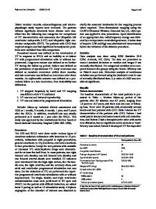
the earliest ventricular activation of a focal VT, during sustained and tolerated arrhythmia, in the proximal middle cardiac vein (pMCV), within 2 cm from the ostium. During the induction of sustained VT, radiofrequency energy (25 W, 65 °C) was delivered in the pMCV, with interruption of VT. Impedance values were monitored throughout the radiofrequency application, to ensure the safety of ablation at this site. The child was discharged without antiarrhythmic therapy and had no relapsing of any arrhythmia as revealed by the implantable loop recorder. During follow-up, the girl perceived a big improvement in her physical abilities even during sports; this fact could let us think that she actually used to experience some limitations in functional capacity due to arrhythmic burden. Our case shows that basal crux VTs can be successfully and safely eliminated with epicardial ablation from MCV in pediatric patients. In these cases, coronary angiography can also be performed to determine proximity to relevant branches of coronary arteries (Figs. 1 and 2).
J Interv Card Electrophysiol
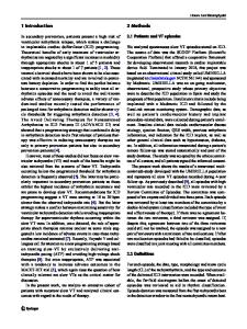
Fig. 1 a Angiography of cardiac venous system. The red dot represents the site of RF delivery. b 3D activation map in LAO and RAO view and ECG with bipolar/unipolar electrograms at the site of the earliest ventricular activation during VT Fig. 2 Real-time tracing during RF ablation. VT termination occurs in 4.2 s of RF delivery
Compliance with ethical standards
References
Conflict of interest The aut
Data Loading...