Visualizing and quantifying cerebrospinal venous flow using PC-VIPR
- PDF / 508,719 Bytes
- 2 Pages / 595.28 x 793.7 pts Page_size
- 89 Downloads / 255 Views
WORKSHOP PRESENTATION
Open Access
Visualizing and quantifying cerebrospinal venous flow using PC-VIPR Eric M Schrauben1*, Kevin Johnson1, Aaron Field2, Oliver Wieben1,2 From 15th Annual SCMR Scientific Sessions Orlando, FL, USA. 2-5 February 2012 Summary Presented is a novel MR imaging protocol to comprehensively assess vessel lumen and hemodynamics of the cerebrospinal veins. The 3 station exam is designed to cover middle cerebral, jugular, and azygous veins in order to investigate the CCSVI hypothesis for the association of insufficient venous return with multiple sclerosis. Background Recently, Zamboni et al. have postulated that chronic cerebrospinal venous insufficiency (CCSVI) with reflux to the level of the deep cerebrospinal veins precipitates increased iron deposition and a neuroinflammatory cascade of events that ultimately result in Multiple Sclerosis (MS) (1). He proposed 5 ultrasound (US) based criteria of diameter changes and impaired flow in the cerebrospinal veins to diagnose MS (2), some of which are hard to assess because they require special US hardware and technologist training and US measures are known to be user-dependent. Here we describe our initial experience with a 4D MR Flow approach, PCVIPR (3), to overcome these problems with a user-independent, non-invasive technique that provides whole vessel coverage. Methods To investigate vessel anatomy and hemodynamics, 7 healthy volunteers were imaged on a clinical 3T system (750 Discovery, GE Healthcare). PC-VIPR of the cerebral, internal jugular (IJV), and azygous veins were performed in a three station exam with a neurovascular phased array and a cardiac coil. Sample imaging parameters for cerebral veins are: Dual Echo, FOV: 24 x 24 x 16 cm, Res: 0.6 x 0.6 x 0.6 mm, 9000 Projections 1 Medical Physics, University of Wisconsin - Madison, Madison, WI, USA Full list of author information is available at the end of the article
(36x), TR=15.9, BW = 31.25, VENC = 40 cm/s, 7:30 min scan time. Acquisitions were modified to allow for retrospective cardiac gating, with additional modifications being investigated for retrospective double-gated cardiac and respiratory reconstruction to address phasic changes in venous flow due to respiration. A single injection of gadofosveset trisodium (Ablavar, Lantheus Medical Imaging, 0.03-0.05 mmol/kg at 3 ml/s) was used for a first pass perfusion scan, contrast-enhanced MRA, and the PC-VIPR scans.
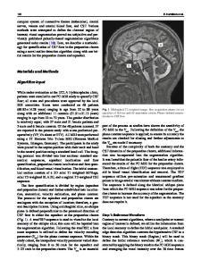
Results Figure 1 shows representative results in the cerebral veins. PC-VIPR allows whole head coverage with high isotropic spatial resolution and visualization of flow characteristics can be collected with retrospective selection of vessels of interest in an arbitrary orientation. Example flow visualizations from the three PC-VIPR sequences are shown in Figure 2. Measurement planes are placed orthogonally to the IJV, from which reflux over the cardiac cycle can be easily detected and quantified. Conclusions This feasibility study demonstrates the potential for 4D MR flow acquisitions to noninvasively measure and visual
Data Loading...