White Paper: Mapping nanomechanical properties of polymers with AFM
- PDF / 892,935 Bytes
- 3 Pages / 585 x 783 pts Page_size
- 107 Downloads / 345 Views
®
CORPORATE PARTNER WHITE PAPER
Mapping nanomechanical properties of polymers with AFM ASYLUM RESEARCH
T
he atomic force microscope (AFM) is an invaluable instrument for characterizing polymer materials at small length scales.1–4 Its spatial resolution enables visualization of submicrometer and subnanometer polymer morphology as well as mapping nanomechanical properties. Mechanical properties of polymers are an important consideration in applications ranging from food packaging to flexible electronics. To optimize mechanical performance, one or more phase-separated components or fillers may be included in polymers. The length scales of such inclusions demand mechanical-property measurements with nanoscale spatial resolution. AFM offers a wide range of techniques for investigating nanomechanical properties, ranging from simple qualitative techniques to more sophisticated quantitative methods. In many cases, these techniques are complementary and can be used together to learn more about polymer samples. Tapping mode has been and still is the most widely used scanning technique, whereby the AFM tip is oscillated above the surface, avoiding sample damage.5 When a phase shift in tapping mode was discovered to yield material property contrast, phase imaging became a source of much excitement beginning in the late 1990s. Since then, phase imaging has become a valuable technique for polymer characterization, where it can often resolve fine structural details and discriminate between various material components. Interpretation is not always straightforward, however, because the Asylum Research The MRS Corporate Partner Program supports the Materials Research Society Foundation.
796
MRS BULLETIN
•
VOLUME 40 • OCTOBER 2015
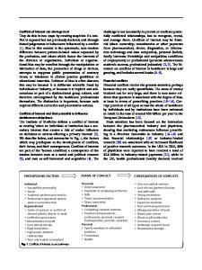
phase response depends on how the material stores elastic energy and dissipates viscous energy (i.e., the loss tangent) as well as other dissipative forces. Notwithstanding these challenges, phase imaging remains a simple and popular means of obtaining qualitative material property contrast (Figure 1). Bimodal imaging (Dual AC)* is another option for qualitative mapping of material property variations. It operates the same as the regular tapping mode with phase imaging, except that
a
an additional resonance mode of the cantilever is driven simultaneously with operation at the first mode. The amplitude and phase response at this second mode is measured along with topography and phase from the first mode. Like regular phase imaging, interpretation of the results is not always easy, but the technique can be useful for obtaining contrast in cases where phase imaging does not provide it, as shown in Figure 2. Most recently, AM-FM Viscoelastic Mapping Mode* has been adopted as the only mode compatible with small cantilevers for fast scanning and is especially well suited for polymers. Similar to bimodal imaging, it uses tapping mode operating simultaneously at two different cantilever mode frequencies. However, the frequency of the second mode is tracked and related to the sample stiffness while the am
Data Loading...