X-Ray microanalysis of phosphorus segregation in type 304L stainless steels
- PDF / 337,753 Bytes
- 3 Pages / 597.6 x 774 pts Page_size
- 80 Downloads / 254 Views
a Al3
= a A12 /V2
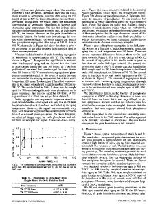
where Al2 = a-Mn, X phase; and A13 = ,B-Mn, a new phase obtained here. Thus, this newly revealed A13-type phase, which we call the'" phase, has a close relation to the X phase. It is suggested that a little change in the solidification condition may determine whether the'" phase or X phase is dominant in the alloy. It is not clear whether the '" phase forms an ordered structure in itself or not. However, the experimental X -ray diffraction pattern extensively fitted the calculated one which was deduced by taking account only of the arrangement of the iron atoms in the unit cell (Figure 4). Furthermore, no additional peak due to ordered structures was detected. Accordingly, it seems that most chromium and molybdenum atoms substitute iron atoms at random in this case. (In some cases, we observed ordered structures altered slightly from the A13 type.[8 J) Carbon atoms may occupy the interstitial sites. From the above results and considerations, it is concluded that the newly revealed phase is essentially a solid solution of the A13 type. The summary of this investigation is that a new phase of the A 13 type was produced in a high-carbon iron alloy containing chromium and molybdenum upon a rapid solidification.
REFERENCES 1. R.C. Ruhl and M. Cohen: TMS-AlME, 1969, vol. 245, pp. 241-51. 2. K.W. Andrews, 0.1. Dyson, and S.R. Keown: Interpretation of Electron Diffraction Patterns, Plenum Press, New York, NY, 1967, p. 180. 3. T. Iwadachi, A. Inoue, T. Minemura, and T. Masumoto: l. lpn. Inst. Met., 1988, vol. 44, pp. 245-54. 4. A. Inouc, L. Amberg, M. Oguchi, U. Backmark, N. Backstrom, and T. Masumoto: Mater. Sci. Eng., 1987, vol. 95, pp. 101-14. 5. K. KishiJake, H. Era, and F. Otsubo: Scripta Metall. Mater., 1990, vol. 24, pp. 1269-73. 6. B.D. Preston: Phil. Mag., 1928, vol. 5, pp. 1207-25. 7. W.B. Pearson: A Handbook of Lattice Spacings and Structures of Metals and Alloys, Pergamon Press, 1958, p. 88. 8. F. Otsubo, E. Tanaka, H. Era, and K. Kishitake: l. Electron Microsc., lpn., 1990, vol. 39, p. 205.
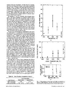
X-Ray Microanalysis of Phosphorus Segregation in Type 304L Stainless Steels E.A. KENIK It has been suggested that intergranular segregation of impurities has an exacerbating influence on the corrosion and high-temperature creep resistance of austenitic stainless steels.[1,2.3J Auger electron spectroscopy (AES) has been employed in many of these studies, especially after the application of hydrogen charging to promote intergranular fracture.[4J On the other hand, X-ray microanalysis in an analytical electron microscope (AEM) can also be used to detect segregation to boundaries. [5-9J A field emission gun AEM can provide higher lateral spatial resolution (approaching 2 nm) in both imaging and X-ray microanalysis, whereas AES generally provides greater sensitivity and spatial resolution perpendicular to the boundary. A recent review of the application of an AEM to segregation, its strengths, and its weaknesses has been given by Williams and Romig.[loJ In this communication, X-ray microanalysis results for
Data Loading...