3D whole heart imaging in severe funnel chest and non-compaction cardiomyopathy
- PDF / 494,512 Bytes
- 2 Pages / 595.276 x 790.866 pts Page_size
- 19 Downloads / 200 Views
IMAGES IN CV APPLICATIONS
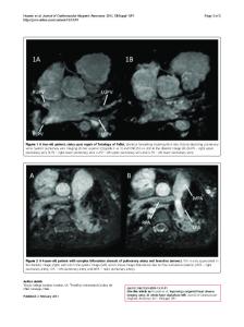
3D whole heart imaging in severe funnel chest and non-compaction cardiomyopathy M. Polacin1,3 · P. Biaggi2 · H. Alkadhi3 · S. Kozerke1 · R. Manka1,3 Received: 11 September 2020 / Accepted: 14 September 2020 © The Author(s) 2020
A 24-year-old female patient without cardiac symptoms underwent preoperative echocardiography before correction of severe pectus excavatum. Echocardiography was difficult to perform because of the funnel chest, nevertheless, hypertrabeculated apical segments were diagnosed. To confirm the suspicion of noncompaction cardiomyopathy (NCCM) cardiac magnetic resonance imaging (MRI) was performed. For better visualization of non-compacted and compacted myocardium of the left ventricle (LV) a 3-dimensional (3D) whole heart sequence (0.9 mm slice thickness, isotropic resolution) covering the entire heart and adjacent thoracic structures was performed. The 3D whole heart sequence revealed a severe funnel chest (Fig. 1a) with Haller-Index of 48 (transverse diameter of the chest divided by the distance between the anterior surface of the vertebral body and the posterior surface of the sternum, normal ratio
Data Loading...