[ 68 Ga]Ga-tilmanocept PET/CT lymphoscintigraphy for sentinel lymph node detection in early-stage oral cavity carcinoma
- PDF / 2,245,854 Bytes
- 2 Pages / 595.276 x 790.866 pts Page_size
- 12 Downloads / 283 Views
IMAGE OF THE MONTH
[68Ga]Ga-tilmanocept PET/CT lymphoscintigraphy for sentinel lymph node detection in early-stage oral cavity carcinoma Rutger Mahieu 1 & Gerard C. Krijger 2 & F.F. Tessa Ververs 3 & Remmert de Roos 2 & Remco de Bree 1 & Bart de Keizer 2 Received: 24 August 2020 / Accepted: 29 September 2020 # The Author(s) 2020
The sentinel lymph node (SLN) procedure is routinely performed for nodal staging in several malignancies, including early-stage oral cancer. In oral cancer, the SLN imaging procedure usually consists of peritumoral injections with a [99mTc]Tc-labelled radiotracer followed by dynamic and planar lymphoscintigraphy and SPECT/ CT [1, 2]. A frequently discussed limitation of this procedure in oral cancer arises in situations where SLNs are located in close vicinity of the radiotracer injection site. Due to the limited resolution of conventional scintigraphy and SPECT/CT, injection site activity can hide adjacent SLNs and hamper discrimination between injection site and SLNs (shine-through phenomenon), potentially resulting in false-negative SLN procedure outcomes [3, 4]. PET/CT lymphoscintigraphy may offer a solution, as it provides superior spatial resolution compared with conventional scintigraphy and SPECT/CT [4, 5]. Here, we present the first within-patient comparison between PET/CT lymphoscintigraphy using [68Ga]Ga-
This article is part of the Topical Collection on Oncology - Head and Neck. * Bart de Keizer [email protected] 1
Department of Head and Neck Surgical Oncology, University Medical Center Utrecht, Utrecht, the Netherlands
2
Department of Radiology and Nuclear Medicine, University Medical Center Utrecht, Heidelberglaan 100, 3584, CX Utrecht, the Netherlands
3
Department of Pharmacy, University Medical Center Utrecht, Utrecht, the Netherlands
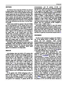
tilmanocept (10 MBq; 15 min post-injection) and SPECT/CT with [99mTc]Tc-tilmanocept (74 MBq; 2 h post-injection) in a cT1N0 tongue cancer patient, both acquired on the day before surgery. Maximum intensity projection images (MIP) of PET (D) demonstrate its superior resolution compared with SPECT (H). Furthermore, two separate lymph vessels can be identified on PET/CT lymphoscintigraphy (D), which are not visualized on SPECT/CT (H). Also note that the activity in a SLN in level Ib on the right site is better visible on axial (A), sagittal (B), and coronal (C) PET/CT lymphoscintigraphic images compared with corresponding SPECT/CT reconstructions (E,F,G). Surgically, five SLNs were localized and harvested (level Ib, 3x level IIa and level III), using a conventional gammaprobe. Histopathological assessment showed metastasis in one SLN located in level IIa. Complementary neck dissection of level I-IV showed no additional lymphatic metastasis (Figure 1).
Eur J Nucl Med Mol Imaging
Figure 1 P[68Ga]Ga-tilmanocept PET/CT vs. [99mTc]Tc-tilmanocept PET/CT.
Funding Open access funding provided by University Medical Center Utrecht.
References 1.
Compliance with ethical standards Conflict of interest The authors declare that they have n
Data Loading...