A simple method for characterizing left ventricular remodeling by cardiovascular magnetic resonance
- PDF / 192,522 Bytes
- 2 Pages / 595.276 x 793.701 pts Page_size
- 109 Downloads / 327 Views
POSTER PRESENTATION
Open Access
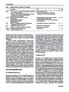
A simple method for characterizing left ventricular remodeling by cardiovascular magnetic resonance Shawn C Pun1*, Maria Figura2, Kelvin Chow2, Mark Haykowsky2, Richard Thompson2, Ian Paterson2 From 2011 SCMR/Euro CMR Joint Scientific Sessions Nice, France. 3-6 February 2011 Background Remodeling of the left ventricle (LV) occurs in response to various physiological and pathophysiological conditions. The American Society of Echocardiography recommend that LV remodeling be described as the relationship between LV mass and LV wall thickness based on 1-D and 2-D measurements. Cardiac magnetic resonance (CMR) provides accurate 3-D measures of LV volumes and mass; however, there is no universally agreed upon approach to measure remodeling. We propose using the ratio of LV mass to LV end-diastolic volume (LVEDV) as the left ventricular remodeling index (LVRI). Objectives To describe and compare patterns of LV remodeling in various cardiac conditions using CMR. We hypothesized that the LVRI would accurately reflect underlying pathophysiology. Methods A total of 105 consecutive cases (89 males, mean age 48±18), with elevated LV mass (referenced to Hudsmith et. al., JCMR 2005) from 2006-2009 were obtained from the CMR clinical database at our institution. 32 healthy volunteers served as controls. Based on the clinical history and the qualitative CMR findings, cases were categorized as: inflammatory (INF), dilated cardiomyopathy (DCM), ischemic cardiomyopathy (ICM), pressure loaded (PL) (eg. aortic stenosis and systemic hypertension) and volume loaded (VL) (eg. aortic regurgitation and mitral regurgitation).
Quantitative volumetric analyses were performed using standard imaging analysis software (Leonardo, Siemens). A short axis stack of steady state free precession cines were used to obtain LV mass and LVEDV by methods of disks. The LVRI was calculated from the ratio of LV mass to LVEDV. Statistical significance was assessed using an unpaired Student’s T-test.
Results The DCM, ICM and PL groups were significantly older and the INF group had a greater proportion of males compared to controls. There was no statistically significant difference in LVRI between males and females or between older (age >35) and younger adults. The mean LVRI for controls was 0.87±0.1g/ml. Compared to controls, mean LVRI was elevated in INF (0.99±0.15g/ml, p=0.002) and PL (0.98±0.12g/ml p=0.002). Conversely, mean LVRI was reduced in ICM (0.79±0.13g/ml, p=0.014) and VL (0.74±0.13g/ml, p2SD control
NA
5 (25%)
12 (35%)
3 (15%)
4 (25%)
5 (33%)
All values are presented as Mean±SD. *P-value 2SD control (proportion of LVRI greater than 2SD above control mean), Mass Index (Mass/BSA).
Author details 1 University of British Columbia, Vancouver, BC, Canada. 2University of Alberta, Edmonton, AB, Canada. Published: 2 February 2011
doi:10.1186/1532-429X-13-S1-P277 Cite this article as: Pun et al.: A simple method for characterizing left ventricular remodeling by cardiovascular magnetic resonance. Journal of Cardiovascu
Data Loading...