Absence of Internal Jugular Vein Flow: a Diagnostic Dilemma
- PDF / 631,349 Bytes
- 3 Pages / 595.276 x 790.866 pts Page_size
- 109 Downloads / 315 Views
CASE REPORT
Absence of Internal Jugular Vein Flow: a Diagnostic Dilemma Rajeshwary Aroor 1
&
Vishwanath Serigar 2 & T M Amulya 1 & Raghuraj Uppoor 3
Received: 30 June 2019 / Accepted: 7 May 2020 # Indian Association of Surgical Oncology 2020
Introduction Developmental venous anomalies are seen in 0.05 to 0.25% of general population [1]. Congenital absence of internal jugular vein is rare. As per our knowledge, very few cases (in single digits) of absent internal jugular vein were reported in English literature. Variability in venous drainage patterns in the head and neck area is important for the head and neck surgeons in order to avoid any intra-operative life-threatening complications. Even the intensivists should be aware of anatomical variations of internal jugular vein prior to central line cannulation.
Case Report A 62-year-old male presented to ENT outpatient department with a mass in the inner aspect of left cheek for 5 months. Patient did not give any history of previous surgeries or previous hospitalizations. Oral cavity examination revealed a 3 × 3 cm ulceroproliferative growth in the left buccal mucosa extending anteriorly corresponding
* Rajeshwary Aroor [email protected] 1
Department of ENT, K S Hegde Medical Academy, Mangalore, India
2
Department of Onco surgery, A J Institute of Medical Sciences Mangalore, Mangalore, India
3
Department of Radiodiagnosis, K S Hegde Medical Academy Mangalore, Mangalore, India
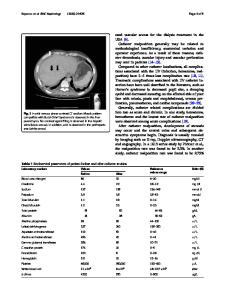
to the lower first premolar tooth posteriorly upto the retromolar trigone. Examination of the neck revealed a 1.5 × 1.5-cm lymph node in left level II region which was mobile and firm in consistency. Biopsy of the primary was suggestive of well-differentiated squamous cell carcinoma. Contrast enhanced computed tomography of neck was showing ipsilateral level II lymph node with absent left internal jugular vein. Radiologically, it was staged as T2N1Mx. Thrombosis of left internal jugular vein due to chronic compression by lymph node was initially thought off (Fig. 1). Since the lymph node was too small and unlikely to cause thrombosis, venous Doppler was done to rule out systemic and cardiothoracic causes for thrombosis and also extension of thrombosis into the thorax. Again, internal jugular vein was not visualized throughout its course. Intra-thoracic venous system was normal in venous Doppler, which again gave a suspicion of external compression. Patient was further subjected to computed tomography venography which showed a narrowed left internal jugular vein at skull base with non-visualization of (thrombosis) of internal jugular vein in the neck and multiple collaterals in the neck and thorax (Fig. 2). Patient underwent wide excision of the primary and nasolabial flap reconstruction of primary defect with left nerve preserving modified neck dissection. During surgery, carotid sheath contained only the carotid artery and its branches and vagus. Internal jugular vein was not seen from the jugular foramen upto the clavicle (Fig. 3). External jugular vein was dilated with
Data Loading...