Diagnostic dilemma in spontaneous innominate artery pathology: a case report
- PDF / 818,391 Bytes
- 4 Pages / 595.276 x 790.866 pts Page_size
- 22 Downloads / 337 Views
CASE REPORT
Diagnostic dilemma in spontaneous innominate artery pathology: a case report Christophoros Kotoulas1 · Charalampos Georgiou1 · Elefterios Chouliaras2 · Nikolaos Theodosiadis1 · Sotirios Kotoulas3 · Athanassios Kotoulas3 · Ioannis Panagiotou1 Received: 25 May 2020 / Accepted: 20 August 2020 © The Japanese Association for Thoracic Surgery 2020
Abstract We report the case of a 71-year-old female who presented with sudden onset of right cervical pain and ipsilateral arm hypoaesthesia. The diagnostic evaluation revealed a pathology of the origin of the innominate artery (IA). The differential diagnosis was among a spontaneous acute dissection and a ruptured pseudoaneurysm. The dilemma of the proper treatment emerged. Surgical or medical treatment? Open or endovascular approach? The patient was offered an open treatment under cardiopulmonary bypass and sort circulatory arrest. As less than ten cases of isolated IA dissection have been previously reported in the literature, we discuss the differential diagnosis difficulties and the treatment options. Keywords Innominate artery · Dissection · Surgery · Aortography · Computed tomography aortography
Introduction Supra-aortic aneurysms are very rare (0.4–4% of all types of aneurysms). Isolated aneurysms of the IA are even rarer, with a reported incidence of 0.003%. [1] A significant percentage is pseudoaneurysms which can be induced by variable etiologies including trauma, iatrogenic insults, and infections. Moreover, less than ten cases of isolated ΙΑ dissection have been previously reported in the literature. [2–9] We present such a case of isolated ΙΑ dissection discussing the differential diagnosis and treatment challenges.
Case report A 71-year-old female presented with sudden onset of right cervical pain and ipsilateral arm hypoaesthesia. She was hemodynamically stable and her medical history was free. * Christophoros Kotoulas [email protected] 1
Cardiac Surgery Department, 401 Army General Hospital of Athens, Kifissias Ave 38, 11526 Athens, Greece
2
Department of Anesthesiology, 401 Army General Hospital of Athens, Athens, Greece
3
Faculty of Medicine, Charles University in Prague, Pilsen, Czechia
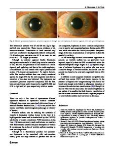
The emergency room laboratory tests were normal. The Doppler scan waveform of the right common carotid artery was slightly damped compared to the left. Computed tomography aortography (CTA) showed a complicated lesion of the orifice of the ΙΑ with a maximum diameter of 2.5 cm and no contribution of the adjusted aorta (Fig. 1a, b). Magnetic resonance angiography (MRA) provided no further information while digital subtraction angiography (DSA) made the diagnosis even more ambiguous showing a small saccular aneurysm of the aortic arch between the IA and the left carotid artery (Fig. 1c). The patient was led to the surgery 32 h after the onset of the symptoms without a definitive diagnosis. With median sternotomy and under cardiopulmonary bypass, the blood supply to the innominate artery branches was restored with an 8 mm ringed polytetrafluoroet
Data Loading...