Analysis of Epithelial Architecture and Planar Spindle Orientation in the Drosophila Wing Disc
The Drosophila melanogaster wing imaginal disc is an epithelial sac that exhibits dramatic tissue growth during the larval stage. With its simple morphology and accessibility of genetic tools, studies using the wing disc have contributed to the understand
- PDF / 611,639 Bytes
- 12 Pages / 504.567 x 720 pts Page_size
- 68 Downloads / 255 Views
Analysis of Epithelial Architecture and Planar Spindle Orientation in the Drosophila Wing Disc Yu-ichiro Nakajima Abstract The Drosophila melanogaster wing imaginal disc is an epithelial sac that exhibits dramatic tissue growth during the larval stage. With its simple morphology and accessibility of genetic tools, studies using the wing disc have contributed to the understanding of the mechanisms of epithelial homeostasis including the control of mitotic spindle orientation. This chapter describes a detailed protocol for analyzing epithelial architecture and planar orientation of the mitotic spindle in the wing disc epithelium. The rapid dissection method, effective immunostaining, and mounting tips described here facilitate genetic and cell biological studies of the wing disc and can be applied to a wide array of studies using various Drosophila tissues. Key words Cell division, Confocal microscopy, Drosophila, Epithelial architecture, Immunostaining, Planar spindle orientation
1
Introduction Epithelial organization is prerequisite for multicellularity, and epithelial tissue is a conserved building block of the metazoan body [1]. Epithelial cells are characterized by apicobasal polarity and intercellular adhesive junctions, which allow tight associations, or communication, with neighboring cells and enable morphogenesis and tissue growth in the layer of epithelial sheet [2]. During development and homeostasis, polarized epithelial architecture is robustly maintained by inherent homeostatic mechanisms [3]. One such cellular mechanism is the control of mitotic spindle orientation during cell division. In proliferating epithelia, the mitotic spindle is aligned parallel to the plane of the epithelium, referred to as planar spindle orientation [4, 5]. Given that planar alignment of the mitotic spindle is predominant in most epithelia and contributes to the maintenance of tissue architecture, understanding its regulatory mechanisms provides insights into the molecular control of epithelial health and disease. The Drosophila wing imaginal disc is a long-favored model for a variety of processes in developmental biology [6, 7]. Structurally, the mature larval wing disc is a flattened epithelial sac composed of
Yu-ichiro Nakajima
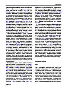
Fig. 1 Schematic images of the Drosophila larval wing imaginal disc. The presumptive wing blade (wing pouch), the presumptive hinge, and the presumptive notum are colored magenta, blue, and orange, respectively. The wing disc is composed of two opposing, continuous epithelial layers: a squamous epithelium, and a pseudostratified epithelium. In the pseudostratified epithelium, cell division occurs at a mitotic zone delimited by apical accumulation of septate junction proteins. At the initiation of mitosis, mitotic nuclei move up in a mitotic zone and the mitotic spindle is aligned to the plane of the epithelium, allowing for planar cell division
apposed pseudostratified and squamous cell monolayers [8] (Fig. 1). In the pseudostratified layer of columnar epithelial cells, polarity and junctional
Data Loading...