Anatomy of the Human Heart
This chapter covers the internal and external anatomy of the heart, its positioning within the thorax, and its basic function. Briefly, the heart is a muscular pump, located in the protective thorax, which serves two functions: (1) collect blood from the
- PDF / 3,570,178 Bytes
- 28 Pages / 595.28 x 790.87 pts Page_size
- 4 Downloads / 313 Views
5
Anthony J. Weinhaus
Abstract
This chapter covers the internal and external anatomy of the heart, its positioning within the thorax, and its basic function. Briefly, the heart is a muscular pump, located in the protective thorax, which serves two functions: (1) collect blood from the tissues of the body and pump it to the lungs and (2) collect blood from the lungs and pump it to all the tissues of the body. The heart’s two upper chambers (or atria) function primarily as collecting chambers, while two lower chambers (ventricles) are much stronger and function to pump blood. The right atrium and ventricle collect blood from the body and pump it to the lungs, and the left atrium and ventricle collect blood from the lungs and pump it throughout the body. There is a one-way flow of blood through the heart which is maintained by a set of four valves (tricuspid, bicuspid, pulmonary, and aortic). The tissues of the heart are supplied with nourishment and oxygen by a separate vascular supply committed only to the heart; the arterial supply to the heart arises from the base of the aorta as the right and left coronary arteries, and the venous drainage is via cardiac veins that return deoxygenated blood to the right atrium. Keywords
Cardiac anatomy • Mediastinum • Pericardium • Atrium • Ventricle • Valves • Coronary artery • Cardiac veins • Cardiac skeleton • Cardiopulmonary circulation
5.1
Introduction
The heart is a muscular pump which serves two functions: (1) collect blood from the tissues of the body and pump it to the lungs and (2) collect blood from the lungs and pump it to all of the tissues of the body. The human heart lies in the protective thorax, posterior to the sternum and costal cartilages, and rests on the superior surface of the diaphragm. The heart assumes an oblique position in the thorax, with two-thirds to the left of midline. It occupies a space between
A.J. Weinhaus, PhD (*) Department of Integrative Biology and Physiology, University of Minnesota, 6-125 Jackson Hall, 321 Church St. SE, Minneapolis, MN 55455-0328, USA e-mail: [email protected]
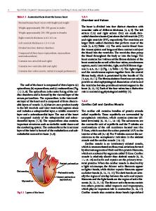
the pleural cavities called the middle mediastinum, defined as the space inside of the pericardium, the covering around the heart. This serous membrane has an inner and an outer layer, with a lubricating fluid in between. The fluid allows the inner visceral pericardium to “glide” against the outer parietal pericardium. The internal anatomy of the heart reveals four chambers composed of cardiac muscle or myocardium. The two upper chambers (or atria) function mainly as collecting chambers; the two lower chambers (ventricles) are much stronger and function to pump blood out of the heart. The role of the right atrium and ventricle is to collect blood from the body and pump it to the lungs. The role of the left atrium and ventricle is to collect blood from the lungs and pump it throughout the body. There is a one-way flow of blood through the heart; this flow is maintained by a set of four valves. The atrioventricular or AV valves (the right tricuspid and left
Data Loading...