Angiographically occult cerebral vascular malformation mimicking cortical venous sinus thrombosis: case report
- PDF / 640,061 Bytes
- 3 Pages / 595.276 x 790.866 pts Page_size
- 38 Downloads / 287 Views
LETTER TO THE EDITOR
Angiographically occult cerebral vascular malformation mimicking cortical venous sinus thrombosis: case report Hongwei Liu1 · Xiaolian Xing1 · Hong Yue1 · Shengwei Gao1 · Weirong Li1 Received: 5 May 2019 / Accepted: 19 September 2019 © Belgian Neurological Society 2019
Keywords Cerebral vascular malformation · Occult · Digital subtraction angiography · Cortical venous sinus thrombosis
Introduction
Case report
Most cerebral vascular malformations (CVMs) are arteriovenous in composition and visible using angiography [1]. Magnetic resonance imaging (MRI) and digital subtraction angiography (DSA) are the gold standard imaging modalities for the evaluation and diagnosis of CVMs [2]. CVMs that are not visible on DSA are known as angiographically occult cerebral vascular malformations (AOVMs) [3]. These include capillary telangiectasias, cavernous angiomas, arteriovenous malformations (AVMs), and intravenous vascular malformations [1]. AOVMs are rare and easily misdiagnosed because of their wide range of clinical manifestations, which include seizures, hemorrhage, headache, and focal neurological deficits [4]. AOVMs that involve hemorrhaging are especially challenging to diagnose. Here, we report a case of angiographically occult AVOM that mimics cortical venous sinus thrombosis (CVST).
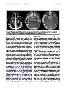
A 31-year-old woman presented with a history of chronically progressive headache and two prior seizures. She was previously diagnosed with polycystic ovary syndrome and had been treated with the oral contraceptive cyproterone acetate for the past 30 months. Her neurological examination was notable for loss of sensitivity to pain and temperature in the right upper and lower limbs. Her D-dimer level was 2.0 mg/L, with a normal coagulation time. Cranial CT scans revealed a hyperdense punctate hemorrhagic lesion in the left frontal lobe (Fig. 1a). T1-weighted images (T1WI), T2-weighted images (T2WI), and susceptibility-weighted MRI images (SWI) showed a large hemorrhagic mass with heterogeneous signal intensity and adjacent edema in the left frontal lobe (Fig. 1b–d). Contrast-enhanced T1-weighted images revealed a partially enhancing lesion with heterogeneous signal (Fig. 1e), and DSA showed no narrow or occlusive venous vascular signal (Fig. 1f). We performed a craniotomy biopsy to differentiate between AVM and CVST. The histopathology of the specimen confirmed an angiographically occult cerebral vascular malformation (cavernous malformation or arteriovenous malformation) (Fig. 1g). The patient was treated with oral contraceptives and antiepileptic agents. We monitored her condition for 6 months, during which she experienced no epileptic episodes. CT scans revealed a region of hypoattenuation during the follow-up examination. We obtained written informed consent from the patient to publish this case report.
* Weirong Li [email protected] Hongwei Liu [email protected] Xiaolian Xing [email protected] Hong Yue [email protected] Shengwei Gao [email protected] 1
Department of Neurology, Taiyuan Central Hos
Data Loading...