Application of field emission scanning electron microscopy for observing irradiated fuel materials
- PDF / 179,964 Bytes
- 4 Pages / 432 x 648 pts Page_size
- 22 Downloads / 343 Views
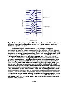
Application of field emission scanning electron microscopy for observing irradiated fuel materials S. Sasaki1, K. Maeda1, A.Yamada2 and T. Asaga1 1 Fuel and Material Department, Oarai Research and Development Center, Japan Atomic Energy Agency, 4002 Narita-cho, O-arai-machi, Higashi-ibaraki-gun, Ibaraki 311-1393, Japan 2 JEOL Ltd, 3-1-2 Musashino, Akishima, Tokyo 196-8558, Japan ABSTRACT The microstructure change of the uranium-plutonium mixed oxide fuels (MOX fuels) irradiated in a fast reactor occurs because of a radial temperature gradient. To make detailed observations and elemental analyses of fuel samples, a field emission scanning electron microscope (FE-SEM) equipped with a wavelength-dispersive X-ray spectrometer (WDX) was installed in a hot laboratory. Because fuel samples have high radioactivities and emit Į-particles, the instrument was modified as follows : 1) The instrument was attached to a remote control air-tight sample transfer unit between a shielded hot cell and the FE-SEM. 2) The FE-SEM was installed in a lead shield box and the control unit was separately located outside the box. After the installation, the microscopy and elemental analyses were applied to low burnup fuel samples. High resolution images were obtained and characteristic X-rays (U, Pu, and so on) emitted from the sample surface were measured. The technique has the great advantage of being able to evaluate the irradiated fuels in detail. In future work, samples of even higher radioactive will be observed and analyzed. INTRODUCTION It is important to study the irradiation behavior of the MOX fuels for design advancement of nuclear reactor fuels. The microstructure change of MOX fuels irradiated in a fast reactor occurs because of a radial temperature gradient[1]-[4]. Typical MOX fuels irradiated in a fast reactor, voids was sweeped towards the fuel center, formation of a central void, and columnar grain structure. The distribution changes of fuel elements and fission product elements occurs during irradiation. Thus, even more detailed observations and elemental analyses of the irradiated MOX fuel surfaces are required than ever before[3] [5] [6]. A FE-SEM, which uses a field emission gun, is capable of imaging at much higher magnification than typical SEM[7]. In addition, FE-SEM has an electron beam current of high stability which is needed for surface elemental analyses. It was necessary to shield operators and devices from radioactivity of samples and to prevent leakage of radioactive materials (especially uranium and plutonium) when irradiated fuel samples are handled for examination. Thus, until now, irradiated fuel samples were handled in hot cells which were air-tight and shielded against radioactivity.
157
In this study, an overview of the installed FE-SEM which was modified for use with irradiated fuel samples and the results of observations and elemental analyses for irradiated fuel samples are reported. EXPERIMENTAL INSTRUMENT The new FE-SEM (JEOL JSM-7001F) was modified and installed in a shielded box. This FE-SEM pro
Data Loading...