Attitudinally Correct Cardiac Anatomy
Anatomy is one of the oldest branches of medicine, dating back as far as the third century bc . Throughout time, the discipline has been served well by a universal system for describing structures based on the anatomic position. Unfortunately, cardiac ana
- PDF / 846,195 Bytes
- 7 Pages / 595.28 x 790.87 pts Page_size
- 87 Downloads / 346 Views
2
Alexander J. Hill
Abstract
Anatomy is one of the oldest branches of medicine, dating back as far as the third century Throughout time, the discipline has been served well by a universal system for describing structures based on the anatomic position. Unfortunately, cardiac anatomy has been a detractor from this long-standing tradition and has commonly been incorrectly described using confusing and inappropriate nomenclature. This is most likely due to the examination of the heart in the Valentine position, in which the heart stands on its apex, as opposed to how it is actually oriented in the body. The description of the major coronary arteries, such as the anterior descending and posterior descending, is attitudinally incorrect; as the heart is oriented in the body, the surfaces are actually superior and inferior. An overview of attitudinally correct human anatomy, the problem areas, and the comparative aspects of attitudinally correct anatomy will be presented in this chapter. BC.
Keywords
Cardiac anatomy • Attitudinally correct nomenclature • Comparative anatomy
2.1
Introduction
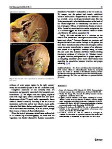
Anatomy is one of the oldest branches of medicine, with historical records dating back at least as far as the third century BC. Cardiac anatomy has been a continually explored topic throughout this time, and there are still publications on new facets of cardiac anatomy being researched and reported today. One of the fundamental tenets of the study of anatomy has been the description of the structure based on the universal orientation, otherwise termed the anatomic position (Fig. 2.1). The anatomic position depicts the subject facing the observer and is then divided into three orthogonal planes. Each plane divides the body or individual structure within A.J. Hill, PhD (*) Medtronic, 8200 Coral Sea Street NE, Mounds View, MN 55112, USA Department of Surgery, University of Minnesota, Minneapolis, MN 55432, USA e-mail: [email protected]
the body (such as the heart) into two portions. Thus, using all three planes, each portion of the anatomy can be localized precisely within the body. These three planes are called (1) the sagittal plane, which divides the body into right and left portions; (2) the coronal plane, which divides the body into anterior and posterior portions; and (3) the transverse plane, which divides the body into superior and inferior portions. Each plane can then be viewed as a slice through a body or organ and will also have specific terms that can be used to define the structures within. If one is looking at a sagittal cut through a body, the observer would describe structures as being anterior or posterior and superior or inferior. On a coronal cut, the structures would be described as superior or inferior and right or left. Finally, on a transverse cut, anterior or posterior and right or left would be used to describe the structures. This terminology should be used regardless of the actual position of the body. For example, assume an observer is looking down at a table and does not move. If a body is lying
Data Loading...