Auer-bodies in cellular components other than typical myeloblasts
- PDF / 558,565 Bytes
- 2 Pages / 595.276 x 790.866 pts Page_size
- 26 Downloads / 274 Views
IMAGES IN HEMATOLOGY
Auer‑bodies in cellular components other than typical myeloblasts Tohru Inaba1 · Taku Tsukamoto2 · Naohisa Fujita1 · Junya Kuroda2 Received: 11 August 2020 / Revised: 2 September 2020 / Accepted: 7 September 2020 © Japanese Society of Hematology 2020
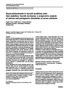
Keywords Auer bodies · Acute myeloid leukemia · Mitotic cells · Large vesicles The Auer body is the hallmark of myeloid neoplasms such as acute myeloid leukemia (AML) or myelodysplastic syndrome. It is typically located in the cytoplasm of neoplastic myeloblasts. In addition, more mature myeloid cells from promyelocytes to segmented neutrophils often contain Auer bodies, especially in patients with acute promyelocytic leukemia (APL) treated with all-trans retinoic acid. However, we encountered a patient with AML in whom Auer bodies were found in other cellular components. A male in his twenties was diagnosed with AML with RUNX1-RUNX1T1 in another hospital. As KIT exon 17 mutation was positive in his leukemic cells, he received cord blood transplantation in his first complete hematological remission. However, 6 months later, he experienced an overt hematological relapse. At that time, his bone marrow was slightly hypercellular, with 90% leukemic blasts. Blasts were large with round to slightly indented fine nuclei, sometimes showing prominent nucleoli. These leukemic blasts sometimes contained fine Auer bodies in their cytoplasm. Notably, fine Auer bodies were also found in mitotic cells and large vesicles suggesting that these cellular components were derived from leukemic cells (Fig. 1).
Theoretically, samples obtained from patients with aggressive hematologic neoplasms such as acute leukemia show mitosis more frequently than those with indolent diseases. However, the origin of mitotic cells is usually not determined using routine May-Giemsa-stained samples. In the literature, only one case report showed the mitotic APL cells which contained several Auer bodies [1]. According to our own experience, extracellular vesicles are sometimes found in hematologic neoplasms such as aggressive lymphoma but seem less frequent in AML. In the review article by Nomura, extracellular vesicles such as large vesicles or oncosomes of 2–50 µm are released by tumor cells [2]. The mechanism of how such large vesicles were produced in this case remains unclarified, but Fig. 1 shows that these large vesicles in his bone marrow were derived from leukemic cells. Though we could not examine precisely, such extracellular vesicles might also contain other disease-specific components such as miRNA derived from the original leukemic blasts, and affect the disease progression.
* Tohru Inaba [email protected]‑m.ac.jp 1
Department of Infection Control and Laboratory Medicine, Kyoto Prefectural University of Medicine, Kawaramachi‑Hirokoji, Kamigyo‑ku, Kyoto 602‑8566, Japan
Division of Hematology and Oncology, Kyoto Prefectural University of Medicine, Kawaramachi‑Hirokoji, Kamigyo‑ku, Kyoto 602‑8566, Japan
2
13
Vol.:(0123456789)
T. Inaba et al.
Fig. 1 Bone m
Data Loading...