Biomechanical Properties of Fibroblasts
- PDF / 3,048,793 Bytes
- 5 Pages / 604.8 x 806.4 pts Page_size
- 45 Downloads / 387 Views
22
were described as a very viscous core bounded by a cortex under persistent tension. 3 , 4 Other eukaryotic cells are more complex, and their mechanical behavior has only recently been investigated. Among them, fibroblasts are a common model system, capable of locomotion and mechanically highly active. In vivo, these cells are embedded in the connective tissue and secrete extracellular components. They can be obtained commercially or can easily be isolated and cultivated in the lab. In the following, we present some state-of-the-art experiments on fibroblasts, leading to a quantitative description of their mechanical behavior. This represents a necessary first step to link biochemistry and biomechanics.
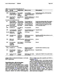
Fibroblasts: Basics In s u s p e n s i o n , fibroblasts have a spherical shape with a diameter of roughly 15 /*m. When allowed to interact
Elastic cortex v
F o l d e d li
Pid membrane
Cytoskeletal filaments Figure 1. The membrane, nucleus, and cytoskeleton are the main structural elements of a fibroblast, which is spherical in suspension.
with a planar substrate in culture conditions, they adhere and spread, maintaining a constant volume during the process. Their apparent surface increases, and membrane wrinkles unfold. Observation from the side reveals that their morphology is close to a truncated sphere. 5 Cell spreading stops when the membrane is taut. Then the fibroblasts exhibit a typical bipolar morphology consisting of a flat lamella at the front and a thin tail at the rear. Under appropriate conditions, cells crawl on their substrate in a cyclic, multistep process/' protrusion at the leading edge in a ruffling fashion, followed and stabilized by adhesion; then cytoskeletal contraction and tail retraction, accompanied by overall structural deformations (Figure 2). In the next sections, we discuss these different aspects of cell mechanics starting with common viscoelastic behavior, then dealing with more original, active processes.
Passive Behavior Fibroblasts behave elastically on the time scale of seconds. This can be made visible by aspirating a sample into a small-diameter micropipet: The fibroblast surface forms a rapid initial protrusion, supposedly linked to the stretching of an actin-rich cortex beneath the plasma membrane. The value of the tension at the surface can be extrapolated to 0.3 nN//xm for the unperturbed cell 5 and increases with an area-expansion modulus of around 10 nN//am upon aspiration. 5,7 Therefore, at rest, the fibroblast surface is only weakly prestrained. A transition from elastic to viscous behavior is clearly seen as the cell starts to slowly creep into the pipet, on a m i n u t e time scale. 5 In addition to a steady flow of cytoplasmic material, this movement requires unfolding of membrane wrinkles. A suitable theoretical model for this situation still needs to be developed, however. Owing to the simple geometry, experiments involving uniaxial pulling of cells between microplates (Figure 3) are easier to interpret.8,9 Using a viscoelastic Kelvin model (a dashpot in
Data Loading...