Canal Wall Down (Radical Cavity)
The posterior canal wall is taken down mainly in cholesteatoma cases when a wide overview of the middle ear structures is necessary. Initially the canal wall is usually preserved to have a landmark for the mastoidectomy. When the canal wall is then taken
- PDF / 188,954 Bytes
- 2 Pages / 439.37 x 666.14 pts Page_size
- 3 Downloads / 323 Views
Canal Wall Down (Radical Cavity)
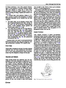
The posterior canal wall is taken down mainly in cholesteatoma cases when a wide overview of the middle ear structures is necessary. Initially the canal wall is usually preserved to have a landmark for the mastoidectomy. When the canal wall is then taken down, drilling is performed parallel to the facial nerve. Once you are medial to the tympanic annulus, it is important to take down the bone overlying the facial nerve (“facial ridge”) as much as possible to allow cleaning and inspection of the middle ear space (Video 5). This is known as lowering the facial ridge and an important step to reduce the incidence of leaving cholesteatoma matrix behind. In this context, also the importance of a wide meatoplasty should be highlighted (Video 6). Once the canal wall is removed, the entrance into the eustachian tube and the canal of the tensor tympani can be seen. The carotid artery lies medial to the eustachian tube (Fig. 8.1).
Landmarks • Horizontal semicircular canal • Tympanic segment of the facial nerve • Head of the malleus • Incudomalleolar joint • Cog • Tensor tympani • Eustachian tube • Supratubal recess • Carotid artery
Electronic supplementary material Supplementary material is available in the online version of this chapter at 10.1007/978-3-70911490-2_8. Videos can also be accessed at http://www.springerimages. com/videos/978-3-7091-1489-6. © Springer-Verlag Wien 2015 C. Arnoldner et al., Manual of Otologic Surgery, DOI 10.1007/978-3-7091-1490-2_8
37
38
8
Canal Wall Down (Radical Cavity)
Fig. 8.1 The posterior canal wall is taken down to gain a wide overview of the middle ear structures (L-SCC lateral semicircular canal, TT tensor tympani, S stapes, asterisk cog, SR supratubal recess, CTT canal of tensor tympani, CA carotid artery, ET eustachian tube)
Data Loading...