Carotid Artery (CA)
Early in the embryogenesis, both primitive proximal ECA and ICA arise separately from the primitive third aortic arch: the ECA from its ventral and the ICA from the dorsal part. The partial involution of the aortic arch, involving its segment distal to th
- PDF / 2,003,546 Bytes
- 17 Pages / 504.57 x 720 pts Page_size
- 78 Downloads / 374 Views
Carotid Artery (CA)
2.1
Cervical Segment
Early in the embryogenesis, both primitive proximal ECA and ICA arise separately from the primitive third aortic arch: the ECA from its ventral and the ICA from the dorsal part. The partial involution of the aortic arch, on both left and right sides, involving its segment distal to the origin of ICA, results in the formation of a common trunk from which develops on each side the common carotid artery (CCA). In the further evolution, the left CCA is annexed by the developped left fourth aortic arch, and the right CCA from the brachiocephalic trunk (Innominate Artery) proximal remnant of the distally completely regressed rigth fourth aortic arch. The common carotid arteries run cranially in the carotid space, surrounded by the three layers of the deep cervical fascia, called the carotid sheet. Approximately at the level of the hyoid bone, usually between the C4 and C6 vertebral bodies, each common carotid artery divides into the internal carotid artery (ICA) and external carotid artery (ECA). Cases of a higher bifurcation, up to the first cervical vertebra (Lie 1968), or lower, in the upper thoracic levels (Vitek and Reaves 1973), have been reported. The carotid sheet is a welldefined structure below the carotid bifurcation, though it is incomplete or absent at the level of the oral–nasal pharynx (Harnsberger 1995). The infrahyoid segment of the carotid space contains the common carotid artery and depending on the level of the bifurcation the proximal part
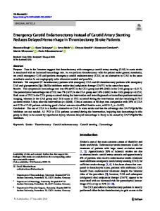
of the ICA, the proximal part of ECA, the Internal Jugular Vein (IJV), portions of the cranial nerves IX, X, XI, XII, the Sympatetic Plexus and Lymph nodes. In the infrahyoid segment, the vessels run in the so-called carotid triangle (Som et al. 2003a) (Fig. 2.1) defined by the sternocleidomastoid muscle, laterally and posteriorly, and by the superior belly of the omohyoid and the posterior belly of the digastric muscle inferiorly and superiorly, respectively. In the suprahyoid–infrahyoid segments, the ICA is accompanied by the IJV located posterolaterally, the cranial nerves (IX, X, XI, and XII), the sympathetic plexus, and the chain of lymph nodes. Near the skull base, the borders of the carotid space (Harnsberger 1995) also called by others (Som and Curtin 2003; Mukherji 2003) the retrostyloid parapharyngeal space can be so outlined: laterally, the parotid space; anteriorly and medially, the parapharyngeal and retropharyngeal spaces, respectively; and posteriorly, the perivertebral space (Fig. 2.2c). The first segment of the ICA (carotid bulb) is slightly enlarged, becoming smaller and narrower 1–2 cm distally. The bulb can be enlarged, particularly in older, atherosclerotic patients, and tortuosity of the distal segment is frequent in very young and older patients. This tortuosity can be congenital or related to dysplastic or atherosclerotic changes. At its origin, the ICA commonly lies posterior and lateral to the ECA. More distally, it is medial to the ECA (Fig. 2.2a, b) (see Chap. 3).
G.B. Bradac, Cerebral Angiog
Data Loading...