Composition and structure of native oxide on silicon by high resolution analytical electron microscopy
- PDF / 1,667,276 Bytes
- 5 Pages / 593.28 x 841.68 pts Page_size
- 62 Downloads / 339 Views
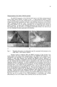
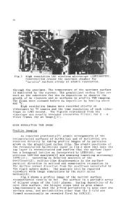
Compositional analysis of thin nanoscale native oxide films formed on {001} silicon wafer surfaces at room temperature was done with an electron energy loss spectrometer coupled to an analytical electron microscope having a field emission source, with better than 4 nm spatial resolution. The electron energy loss spectra show a shift in the threshold onset energy of the Si-L edge of the native oxide from —99 eV loss corresponding to pure elemental silicon to —105 eV loss, and elemental analysis using the ionization regions of the core loss edges showed the composition to be SiO, within a few percent. Microdiffraction and high resolution electron microscopy (HREM) results showed that the native oxide was completely amorphous, and did not contain detectable nanocrystals. The native oxide can be removed from the Si surface by heating in UHV for a short time at 1000 °C. However, this procedure resulted in the formation of small amounts of a crystalline phase on the Si wafer surface, which was shown to be /3-SiC by the same methods.
I. INTRODUCTION
It is well known that a thin layer of material called "native oxide" begins to form immediately on chemically clean silicon surfaces when they are exposed to air at room temperature. The composition and structure of this film have been of interest for many years. Various experimental results have suggested that the native oxide has a composition of SiO* with x < 2.1"3 Electron spectroscopy for chemical analysis (ESCA)1 showed that the native oxides do not have the same composition as thermal oxide Si which is SiO2. Vacuum ultraviolet reflectance spectroscopy (VUVRS)2 results indicated that the native oxide may be a composite of SiO-SiCv Electron energy loss spectroscopy3 results indicated qualitatively that the oxygen content of the native oxide was between Si2O and SiO2 on the basis of study of the low loss region. Recent developments in transmission electron microscopy, notably parallel electron energy loss spectrometers fitted to analytical electron microscopes with field emission sources, and high resolution imaging, enabled direct determination of the structure and composition of these films, using cross section specimen geometry and suitable protection for the films during specimen preparation. In this paper we report the first quantitative electron energy loss spectroscopy (EELS) nanoanalysis of native oxide on silicon using core edges, and the first nanoscale observations of its structure.
II. MATERIALS AND EXPERIMENTAL METHODS Czochralski-grown Si {001} wafers were cleaned in HF at room temperature, rinsed in doubly de-ionized J. Mater. Res., Vol. 5, No. 2, Feb 1990
http://journals.cambridge.org
Downloaded: 14 Mar 2015
water, and then exposed to air to permit native oxide formation until placed in an ultra high vacuum (UHV) chamber. A thin Au film was deposited on the specimen surfaces in the UHV chamber, so that the native oxide layer formed was protected between the Si substrate and the deposited Au film. Specimens were generally held at room temperature durin
Data Loading...