Cytoplasmic VDR expression as an independent risk factor for ovarian cancer
- PDF / 1,359,331 Bytes
- 9 Pages / 595.276 x 790.866 pts Page_size
- 8 Downloads / 254 Views
ORIGINAL PAPER
Cytoplasmic VDR expression as an independent risk factor for ovarian cancer Bastian Czogalla1 · Eileen Deuster1 · Yue Liao1 · Doris Mayr2 · Elisa Schmoeckel2 · Cornelia Sattler1 · Thomas Kolben1 · Anna Hester1 · Sophie Fürst1 · Alexander Burges1 · Sven Mahner1 · Udo Jeschke1,3 · Fabian Trillsch1 Accepted: 13 June 2020 © The Author(s) 2020
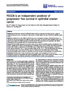
Abstract The vitamin D receptor (VDR), primarily known as a crucial mediator of calcium homeostasis and metabolism, has been shown to play a significant role in various cancer entities. Previous studies have focused on vitamin D and its receptor in gynecological cancers, noting that the receptor is upregulated in epithelial ovarian cancer (EOC). The aim of this study is to analyze the prognostic impact of VDR and its functional significance in ovarian cancer. Through immunohistochemistry, VDR staining was examined in 156 ovarian cancer samples. Evaluation of VDR staining was conducted in the nucleus and the cytoplasm using the semi-quantitative immunoreactive score, and the scores were classified into high- and low-level expressions. Expression levels were correlated with clinical and pathological parameters as well as with overall survival to assess for prognostic impact. Differences in cytoplasmic VDR expression were identified between the histological subtypes (p = 0.001). Serous, clear cell, and endometrioid subtypes showed the highest staining, while the mucinous subtype showed the lowest. Cytoplasmic VDR correlated with higher FIGO stage (p = 0.013; Cc = 0.203), positive lymph node status (p = 0.023; Cc = 0.236), high-grade serous histology (p = 0.000; Cc = 0.298) and grading from the distinct histological subtypes (p = 0.006; Cc = − 0.225). Nuclear VDR did not correlate with clinicopathological data. High cytoplasmic expression of VDR was associated with impaired overall survival (HR 2.218, 32.5 months vs. median not reached; p 60 years
n
Percentage (%)
110 12 21 13
70.5 7.7 13.5 8.3
1 40 18 97
0.6 25.6 11.5 62.3
61 43 52
39.1 27.6 33.3
147 3 6
94.2 1.9 3.8
24 80
21.8 72.7
6 5 8
28.6 23.8 38.1
6 6 0
46.2 46.2 0
12
100.0
35 10 103 3
22.4 6.4 66.0 1.9
83 73
53.2 46.8
Histochemistry and Cell Biology
Ethical approval
Staining evaluation
The Ethics Committee of the Ludwig-Maximilians-University, Munich, Germany, approved this study (approval numbers 227-09 and 18-392). All tissue samples utilized for this investigation were collected from material from the archives of the Department of Obstetrics and Gynecology, University Hospital, LMU Munich, Munich, Germany, having initially been used for pathological diagnostics. The diagnostic procedures were concluded before the current study was conducted. During the analysis, the observers were fully blinded for patients’ data.
Specific VDR immunohistochemically staining reaction was observed in the nuclei and cytoplasm of the cells. The strength and distribution pattern of VDR staining was evaluated using the semi-quantitative immunoreactive score (IR score, Remmele’s score). To obtain th
Data Loading...