Dark cortical rim: an MRI feature of polycystic ovarian syndrome
- PDF / 1,144,871 Bytes
- 9 Pages / 595.276 x 790.866 pts Page_size
- 34 Downloads / 288 Views
PELVIS
Dark cortical rim: an MRI feature of polycystic ovarian syndrome Arwa Badeeb1 · Alexander Brook2 · Karen S. Lee2 Received: 1 July 2020 / Revised: 23 August 2020 / Accepted: 3 September 2020 © Springer Science+Business Media, LLC, part of Springer Nature 2020
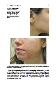
Abstract Purpose To determine if the presence of a dark cortical rim around the ovary on magnetic resonance imaging (MRI) is associated with polycystic ovarian syndrome (PCOS). Materials and methods This retrospective study included 52 PCOS patients with 98 total ovaries and 52 age-matched controls with 104 total ovaries. The ovaries were evaluated on MRI with at least two orthogonal views on T2-weighted sequences. Ovarian volume and follicular count per ovary were measured. Each ovary was also assessed for a dark cortical rim around the ovary on T2-weighted imaging which involved equal to or more than 50% of the ovarian circumference. The degree of rim continuity was classified as continuous if the rim involved greater than 75% of the ovarian circumference, discontinuous if 50–75% of the ovarian circumference was covered, or absent if less than 50% of the ovarian circumference was involved. The rim thickness was measured if present. T test and χ2 tests were performed to compare continuous and categorical variables, correspondingly, between cases and controls. ROC curves and area under the curve (AUC) were used to assess predictive performance and DeLong’s paired test was used to compare AUCs. Results A higher percentage of PCOS patients exhibited a continuous cortical rim about the ovary (71%) and a lower percentage of an absent cortical rim (8%) compared to controls (25% and 37%, respectively) (p
Data Loading...