Detection of the common origin of the radiculomedullary artery with the feeder of spinal dural arteriovenous fistula usi
- PDF / 1,333,566 Bytes
- 8 Pages / 595.276 x 790.866 pts Page_size
- 15 Downloads / 256 Views
SPINAL NEURORADIOLOGY
Detection of the common origin of the radiculomedullary artery with the feeder of spinal dural arteriovenous fistula using slab maximum intensity projection image Masafumi Hiramatsu 1 & Kenji Sugiu 1 & Takao Yasuhara 1 & Tomohito Hishikawa 1 & Jun Haruma 1 Yu Takahashi 1 & Satoshi Murai 1 & Kazuhiko Nishi 1 & Yoko Yamaoka 1 & Isao Date 1
&
Received: 9 March 2020 / Accepted: 22 May 2020 # Springer-Verlag GmbH Germany, part of Springer Nature 2020
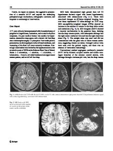
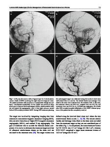
Abstract Purpose Endovascular therapy to the spinal dural arteriovenous fistula (SDAVF) with a common origin of the radiculomedullary artery and the feeder of the shunt has the risk of spinal cord infarction. This study aimed to retrospectively assess the detection rate of normal spinal arteries from the feeder of SDAVF. Methods We retrospectively collected the angiographic and clinical data of SDAVFs. This study included 19 patients with 20 SDAVF lesions admitted to our department between January 2007 and December 2018. We assessed the detection rate of normal radiculomedullary artery branched from the feeder of SDAVF between the period using the image intensifier (II) and flat panel detector (FPD) and evaluated the treatment results. Results The detection rates of the radiculomedullary artery branched from the feeder of SDAVF were 10% (1/10 lesions) during the II period and 30% (3/10 lesions) during the FPD period. During the FPD period, all normal radiculomedullary arteries branched from the feeder were only detected on slab maximum intensity projection (MIP) images of rotational angiography, and we could not detect them in 2D or 3D digital subtraction angiography. All lesions that had a common origin of a normal radiculomedullary artery and the feeder were completely obliterated without complications. There was no recurrence during the follow-up period. Conclusions The flat panel detector and slab MIP images seem to show the common origin of the normal radiculomedullary arteries from the feeder more accurately. With detailed analyses, SDAVF can be safety treated. Keywords Slab MIP image . Spinal dural arteriovenous fistula . Radiculomedullary artery . Spinal artery
Abbreviations AKA Adamkiewicz’s artery ARMedA anterior radiculomedullary artery ASA anterior spinal artery EVT endovascular therapy
Presentation at meeting The abstract of this article was presented in whole at the 15th congress of World Federation of Interventional and Therapeutic Neuroradiology. * Masafumi Hiramatsu [email protected] 1
Department of Neurological Surgery, Okayama University Graduate School of Medicine, Dentistry and Pharmaceutical Sciences, 2-5-1 Shikata-cho, Kita-ku, Okayama 700-8558, Japan
FPD II PRMedA NBCA PSA SDAVF 2D-DSA 3D-DSA
flat panel detector image intensifier posterior radiculomedullary artery n-butyl-cyanoacrylate posterolateral spinal artery spinal dural arteriovenous fistula two-dimensional DSA three-dimensional DSA
Background Spinal dural arteriovenous fistulas (SDAVFs) are rare with an assumed annual incidence of 5–10 cases per one mi
Data Loading...