Electron Spin Relaxation of Tb 3+ and Tm 3+ Ions
- PDF / 1,114,982 Bytes
- 16 Pages / 439.37 x 666.142 pts Page_size
- 17 Downloads / 374 Views
Applied Magnetic Resonance
ORIGINAL PAPER
Electron Spin Relaxation of Tb3+ and Tm3+ Ions Joseph McPeak1 · Dinu Alexander2 · Cyriac Joseph2 · Sandra S. Eaton1 · Gareth R. Eaton1 Received: 17 July 2020 / Revised: 31 August 2020 © Springer-Verlag GmbH Austria, part of Springer Nature 2020
Abstract Electron spin relaxation times T1 and Tm of Tb3+ and Tm3+ in 1:1 water:ethanol and of Tb3+ doped (2%) in crystalline La2(oxalate)3 decahydrate were measured between about 4.2 and 10 K. Both cations are non-Kramers ions and have J = 6 ground states. Echo-detected spectra are compared with CW spectra and with field-stepped directdetected EPR spectra. Due to the strong temperature dependence of T1, measurements were not made above 10 K. Between about 4.2 and 6 K T1 is strongly concentration dependent between 1 and ~ 50 mM. T1 values at 4.2 K are in the µs range which is orders of magnitude faster than for 3d transition metals. Phase memory times, Tm, are less than 500 ns, which is short relative to values observed for 3d transition metals and organic radicals at 4 K. Tm is longer in the oxalate lattice which is attributed to the lower proton concentration in oxalate than in the organic solvent, which decreases nuclear spin diffusion. The rigidity of the crystalline lattice also may contribute to longer Tm.
1 Introduction Lanthanides are of on-going interest because of their wide range of optical and magnetic properties, possible applications in biomolecular structural studies, and unique uses in quantum devices [1–3]. Although lanthanides have been studied extensively by CW EPR, there are a few reports of pulse measurements of electron spin relaxation times, which are fundamental to the performance of some devices. In addition, most prior studies of lanthanides have been in single crystals, and it is of interest to see what information can be obtained from glassy samples. Two ions with non-Kramers J = 6 ground states, T b3+ and T m3+, were selected for comparisons, because there is little known about the relaxation times of these ions. * Gareth R. Eaton [email protected] 1
Department of Chemistry and Biochemistry, University of Denver, 2101 E. Wesley Ave, Denver, CO 80208, USA
2
School of Pure and Applied Physics, Mahatma Gandhi University, Kottayam, India
13
Vol.:(0123456789)
J. McPeak et al.
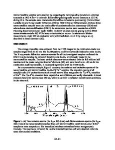
The Tb3+ ion has a 4f8 electron configuration and S = 3. The term with the lowest energy is 7F with L = 3. Strong spin–orbit coupling results in a J = 6 ground state. The full energy-level diagram is shown in Fig. 11.1 of [4]. Tm3+ has a 4 f12 electron configuration with S = 1, L = 5, and the ground state, 3H6, has J = 6. For both ions, the splitting of the MJ levels is symmetry-dependent, but for axial symmetries, MJ = ± 6 is the ground state. These 3+ cations are non-Kramers ions, so to first approximation, one does not expect to observe EPR spectra in octahedral sites with the usual excitation mode in which B1 is perpendicular to B0. It is predicted that the resonance of a non-Kramers ion could be observ
Data Loading...