Extracting elastic properties and prestress of a cell using atomic force microscopy
- PDF / 521,454 Bytes
- 5 Pages / 584.957 x 782.986 pts Page_size
- 26 Downloads / 319 Views
Y.W. Zhang Department of Materials Science and Engineering, National University of Singapore, Singapore 119260 (Received 28 May 2008; accepted 25 November 2008)
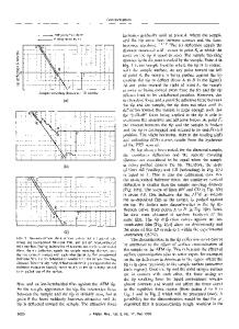
An analytical solution was derived for the indentation of a cell using atomic force microscopy. It was found that the contribution of the cell membrane to the overall indentation stiffness is dependent on the size of the indenter. When a small indenter [for example, an atomic force microscopy (AFM) tip] is used to probe the mechanical properties of cells, the cell membrane and its prestress were important in interpreting indentation data. The solution allows the partition of contributions from the membrane and the interior soft phase. The apparent elastic modulus of the cell and the prestress of the cell membrane can be extracted. In addition, the modulus of the cell membrane could be estimated from the extracted apparent modulus if the interior soft phase of the cell was known and vice versa. However, when a large indenter is used (for example, a microbead attached to the cantilever beam of the AFM), the contribution of the cell membrane is negligible.
I. INTRODUCTION
The mechanical properties of living cells play an important role in the regulation of their shape changes during essential biological processes such as migration, contraction, morphological changes, crawling, and even alterations in gene expression and protein synthesis.1 In recent decades, atomic force microscope (AFM2) has been widely used in studying the mechanical properties of cells: by pushing the sample against the AFM tip to a certain depth of indentation, a force-indentation curve can be collected, from which mechanical properties of a cell can be determined.3–8 The Hertz model9,10 and Sneddon’s solution11 are the most commonly used approaches to interpret the indentation data. By using such approaches, researchers were able to model the cells as homogeneous elastic half-space although the actual cell is covered by a phospholipid bilayer (cell membrane) usually with nonzero isotropic prestress due to the adhesion to extracellular matrix7 or due to the intrinsic residual stress (for example, cells of heart).12 Many experimental results have shown that the correlation between these models and the force-indentation data was unsatisfactory.13–15 One of the main reasons is that these models do not consider the biphasic structure of cells and the prestress of cell membranes. As a consequence, only an apparent modulus can be determined.
II. METHODS
In the present treatment, the cell membrane was modeled as a pretensed elastic shell with a thickness of h, and the interior phase of the cell (the cytoplasm, the
a)
Address all correspondence to this author. e-mail: [email protected] DOI: 10.1557/JMR.2009.0121 J. Mater. Res., Vol. 24, No. 3, Mar 2009
http://journals.cambridge.org
Therefore, the relative contributions from the cell membrane (with the prestress) and from the interior soft phase to the overall resistance to external mechanical stimuli are unclear. The objective of present
Data Loading...