Giant retroperitoneal abscess with left lower limb extension from perforated colon cancer
- PDF / 1,470,563 Bytes
- 3 Pages / 595.276 x 790.866 pts Page_size
- 65 Downloads / 288 Views
THE LAST IMAGE
Giant retroperitoneal abscess with left lower limb extension from perforated colon cancer R. V. Pandini1 · V. E. Seid1 · L. S. Gerbasi1 · M. N. Figuereiredo1 · A. S. Portilho1 · M. Marcelino1 · S. E. A. Araújo1,2 Received: 19 July 2020 / Accepted: 3 September 2020 © Springer Nature Switzerland AG 2020
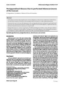
These images show a 63-year-old female, with a history of hypertension. She presented with a perforated adenocarcinoma of the descending colon, with liver metastasis and an unusual large retroperitoneal abscess extending through the extraperitoneal pelvic space and to the left lower limb. This patient underwent a hygienic open left colectomy and a left thigh incision to drain the abscess. Computed tomographyguided drainage was performed 10 days after surgery for complete drainage of the pelvic extraperitoneal abscess. The patient was discharged after 16 days of broad-spectrum antibiotics with complete resolution of sepsis (Figs. 1, 2, 3, 4, 5, 6, 7, 8, 9).
Fig. 2 Coronal computed tomography image of the retroperitoneal abscess and a pelvic extraperitoneal abscess (blue arrows) due to a perforated tumor on the descending colon
Fig. 1 Computed tomography image showing a large descending colon adenocarcinoma (blue circle) perforated to the retroperitoneal space with the surrounding abscess (blue arrows)
* R. V. Pandini [email protected] 1
Colorectal Department, Hospital Israelita Albert Einstein, Av. Albert Einstein 627/701, São Paulo, SP 05652‐900, Brazil
Oncology Department, Hospital Israelita Albert Einstein, São Paulo, SP, Brazil
2
Fig. 3 Sagittal computed tomography image of the retroperitoneal abscess and its ascending and descending pathway
13
Vol.:(0123456789)
Techniques in Coloproctology
Fig. 4 Computed tomography image of the abscess trajectory to the left thigh
Fig. 6 The left colectomy specimen and the perforated tumor (blue circle)
Fig. 5 Image after left colectomy showing the retroperitoneal abscess cavity due to the tumor perforation, its cranial relation to the kidney and the retroperitoneal structures
Fig. 7 Left thigh edema and inflammation due to the abscess
13
Techniques in Coloproctology
Compliance with ethical standards Conflict of interest The authors have no potential conflict of interest. Ethical approval This case was conducted in line with the ethics committee and instructions of Hospital Municipal Vila Santa Catarina. Informed consent The participant has consented to the submission of the case report to the journal.
Publisher’s Note Springer Nature remains neutral with regard to jurisdictional claims in published maps and institutional affiliations.
Fig. 8 An 8-cm left thigh incision made to drain the abscess
Fig. 9 Postoperative pigtail drainage of extraperitoneal abscess
13
Data Loading...