Periappendiceal abscess due to perforated adenocarcinoma of the caecum
- PDF / 1,102,728 Bytes
- 5 Pages / 595.276 x 793.701 pts Page_size
- 91 Downloads / 312 Views
CASE REPORT
Periappendiceal Abscess Due to perforated Adenocarcinoma of the Caecum G. Fragandreas, E. Synekidou, A. Marneri, N. Tona, M. Verroiwtou
Abstract Colon adenocarcinomas rank among the most common malignancies. Characteristic complications include obstruction and chronic anaemia. Perforation with consequent abscess formation is a rather atypical presentation. We present the case of a 62-year-old male patient with a periappendiceal abscess resulting from perforated caecal carcinoma. Since clinical and intraoperative findings were suggestive of associated malignancy, a right hemicolectomy with latero-lateral ileotransverse anastomosis was performed. Periappendiceal mass is a common surgical entity. The differential diagnosis includes a variety of benign conditions (infectious or inflammatory) and neoplasia. The diagnosis of associated malignancy, when radiological examination is not diagnostic, requires a high index of suspicion in order to provide the appropriate treatment. Key words: appendiceal mass, periappendiceal abscess, colorectal cancer, cecal carcinoma
Introduction Colorectal cancer is one of the most common malignancies worldwide. Acute appendicitis is one of the most frequent causes of acute abdomen at all ages. The association of these two entities was described for the first time more than a century ago. Nonetheless, the diagnostic and therapeutic approach remains a challenge, especially when dealing with a periappendiceal mass. There is always a risk of misdiagnosing a malignancy and not offering the patient adequate treatment. The surgeon should always be suspicious of this association, particularly in complicated cases, and even in younger patients. A radical resection is the most appropriate treatment for associated malignancy.
Case Report A 62-year-old diabetic male patient was referred to our department with a two-week history of fever reaching 38°C, constipation, vomiting and right lower quadrant abdominal pain. He reported a weight loss of five kilograms in the last six months. Clinical examination revealed normal bowel sounds on auscultation and a right lower quadrant G. Fragandreas, E. Synekidou, A. Marneri, N.Tona, M. Verroiwtou A' Surgical Department of Hippokration General Hospital of Thessaloniki, Greece Corresponding author: Eirini Synekidou Md, Anatolikis Thrakis 2, 54453 Thessaloniki, Greece. (Home address), A' Surgical Department of Hippokration General Hospital of Thessaloniki, Konstantinoupoleos 49, Greece Tel: +00306987177126, +00302310939598 e-mail: [email protected], [email protected] Received 27 June 2014; Accepted 2 August 2014
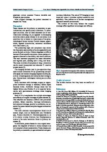
Hellenic Journal of Surgery 86
tenderness with rebound. Digital rectal examination was unremarkable. Laboratory data showed a mild leukocytosis (15.000/mm3) and anaemia (haematocrit 29%, haemoglobin 9.0 g/dl). Parenteral antimicrobial therapy was administered. An X-ray of the abdomen showed small air-fluid levels. Abdominal computed tomography (CT) revealed a mass in the right iliac fossa close to the caecum and the distal ileum
Data Loading...