High-dimensional and high-resolution x-ray tomography for energy materials science
- PDF / 3,931,888 Bytes
- 7 Pages / 585 x 783 pts Page_size
- 12 Downloads / 287 Views
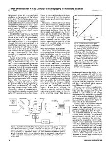
Introduction X-rays have been used for imaging ever since their discovery more than a century ago. Their excellent penetration capability facilitates the reconstruction of the sample’s internal structure noninvasively, especially through the tomography modality, in which the transmission images of the specimen are recorded in different viewing angles. The raw data of a tomographic scan are three-dimensional (3D) (x, y, and θ, which needs to go through a numerical reconstruction procedure that converts it into the 3D Cartesian coordinate system [x, y, and z]).1 Such a numerical reconstruction procedure avoids any ambiguity in the imaging data interpretation caused by “shadows” of features at different depths overlapping in the projective image in the beam direction. The tomography technique has been broadly adopted in many research and clinical applications. Depending on the configuration of the x-ray imaging setup, x-ray tomography can cover a broad range of length scales, provide different contrast mechanisms, and even capture dynamically evolving features of interest (fourdimensional tomography). The implementation of x-ray tomography at modern synchrotron facilities has further opened vast scientific opportunities. A synchrotron source typically delivers x-ray beams with
intensity more than three orders of magnitude higher than a tabletop x-ray source.2 The temporal resolution (often regarded as the fourth dimension of the imaging data) of a tomographic scan is, therefore, dramatically improved to the seconds-level for a microtomography beamline3 and to the minutes level for a nanotomography beamline.4 Another major favorable characteristic of a synchrotron x-ray source is its energy tunability over a very broad range. By conducting tomography at selected energy levels, compositional and chemical sensitivity can be achieved through analyzing the material’s energy-dependent responses.5–9 This experimental strategy is termed spectrotomography and is conceptually similar to the iodine k-edge imaging (i.e., taking images using x-rays above and below the k-edge of iodine contrast agent), which has been broadly used in medical/ clinical applications.10 The use of synchrotron radiation, however, brings it to the next level by offering the capability to probe the valence state’s spatial variation and dynamic evolution for the element(s) of interest at nanoscale resolution. The energy-resolved tomographic scan can also be viewed as a spatially resolved spectroscopic measurement, where each spectrum is associated with a unique volume unit (i.e., a voxel). It therefore fingerprints the local compositional and chemical
Zhenjiang Yu, Department of Electrochemistry, Harbin Institute of Technology, China; [email protected] Jiajun Wang, Harbin Institute of Technology, China; [email protected] Yijin Liu, Stanford Synchrotron Radiation Lightsource, SLAC National Accelerator Laboratory, USA; [email protected] doi:10.1557/mrs.2020.86
• VOLUME © 2020 Materials Research Society MRS BULLETIN 45 •available APRIL 202
Data Loading...