Huge thrombus in the ascending aorta: a case report and literature review
- PDF / 4,354,217 Bytes
- 4 Pages / 595.276 x 790.866 pts Page_size
- 20 Downloads / 246 Views
(2019) 14:157
CASE REPORT
Open Access
Huge thrombus in the ascending aorta: a case report and literature review Baogang Wang, Dashi Ma, Dianbo Cao and Xiaxia Man*
Abstract Background: A floating thrombus in the ascending aorta is occasionally found in clinical practice. The treatment for such lesions is poorly defined and mainly depends on the clinical experience of the surgeons. Case presentation: We herein report a case involving a 22- × 22- × 45-mm space-occupying lesion in the ascending aorta. The patient was successfully treated with surgical intervention. Thrombectomy and ascending aorta replacement were performed to prevent systemic embolization. Histopathological examination revealed that the lesion was a thrombus. Conclusions: Aortic computed tomography angiography is a useful examination technique for patients with aortic thrombi. Resection of the thrombus can effectively reduce the risk of recurrent embolism. Keywords: Floating thrombus, Ascending aorta, Transthoracic echocardiography, Computed tomography angiography
Background A floating thrombus in the ascending aorta is occasionally found in clinical practice. The treatment for such lesions is poorly defined and mainly depends on the clinical experience of the surgeons. Considering the catastrophic effects of embolism, emergency surgery is usually needed. The ethics committee of the Jilin University First Hospital approved this study and the publication of this case report. Informed consent was obtained from the patient. Case presentation A 56-year-old, 61-kg, 168-cm man was admitted to our institution for a routine physical examination. His vital signs were as follows: pulse, 100 beats/min, blood pressure, 140/100 mmHg; oxygen saturation, 95% on room air; and body temperature, 36.5 °C. No murmurs, dyskinesia or paresthesia were detected. The patient had no history of atrial fibrillation. His serum N-terminal pro-brain natriuretic peptide concentration was slightly elevated (131 pg/ml), and his
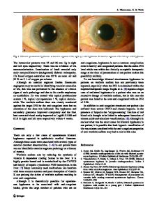
prothrombin time was 10 s. No abnormalities were found in the complete blood count or tumor markers. The serum procalcitonin level was 0.40 ng/ml, the cardiac troponin I level was 0.012 ng/ml, the erythrocyte sedimentation rate was 12 mm/h, and the C-reactive protein level was 3.10 mg/L. A blood culture was negative. Transthoracic echocardiography (TTE) and computed tomography angiography (CTA) revealed a well-defined pedunculated mass measuring 22 × 22 × 45 mm in ascending aortic lumen (Fig. 1a). Magnetic resonance imaging showed multiple asymptomatic cerebral infractions. No abnormalities were found on color Doppler ultrasound of the lower extremities. Furthermore, magnetic resonance imaging demonstrated atherosclerosis of the ascending aorta, and the mass was suspected to be thrombus (Fig. 1b). The patient underwent a thrombectomy. Cardiopulmonary bypass was established by cannulation of the right femoral artery and right atrium. The ascending aorta was clamped proximal to the brachiocephalic trunk. A thrombus that was attached to the aortic
* Corresp
Data Loading...