Impact of ascending aorta replacement by graft on elastic properties of descending thoracic aorta evaluated by cardiac m
- PDF / 1,069,778 Bytes
- 7 Pages / 595.276 x 790.866 pts Page_size
- 82 Downloads / 329 Views
RESEARCH ARTICLE
Impact of ascending aorta replacement by graft on elastic properties of descending thoracic aorta evaluated by cardiac magnetic resonance imaging Marie‑Catherine Morgant1,2 · Johel Miteran2 · Siyu Lin2 · Aline Laubriet1 · Alexandre Cochet2,3 · Alain Lalande2,3 · Olivier Bouchot1,2 Received: 7 March 2019 / Revised: 20 December 2019 / Accepted: 12 January 2020 © European Society for Magnetic Resonance in Medicine and Biology (ESMRMB) 2020
Abstract Objective The aim of our study was to evaluate the impact of aortic root replacement by graft on the elastic properties of the descending thoracic aorta using cardiac magnetic resonance imaging (MRI) and automatic post-processing. Materials and methods Nineteen patients were operated for an aortic root aneurysm. Cardiac MRI was performed before and after surgery to measure aortic compliance. Images were acquired on a 1.5 T MRI with a conventional aortic MRI protocol plus one additional kinetic sequence orientated perpendicularly to the aorta at the level of pulmonary trunk. The contours of the ascending and descending aortas were detected automatically for each phase with homemade software. Results Mean time between surgical procedure and earliest post-operative MRI was 18.2 ± 7.1 months. There was no significant difference between the pre- and earliest post-operative mean descending aorta areas and no significant modification in descending aortic compliance after aortic root replacement (1.43 ± 0.84 vs 1.37 ± 0.58 mm2/mmHg, p = 0.47). Pre- and post-operative systolic and diastolic blood pressure were similar. There was a significant decrease in ascending aortic compliance after surgery (2.52 ± 1.24 vs 0.91 ± 0.52 mm2/mmHg; p 30) Current smoking Connective tissue disorders Bicuspid valve Aortic insufficiency > 2 Sinus of Valsalva, mean diameter (mm)
19 17 55.3 ± 16.3 56.8 ± 16.3 9 4 2 2 3 7 50.1 ± 5.1
89.5%
47.4% 21.1% 10.5% 10.5% 15.8% 36.8%
BMI body mass index, SD standard deviation
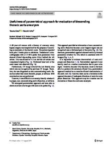
at the level of the bifurcation of the pulmonary artery and during a short breath-hold. If necessary, the image acquisition was repeated to be also perpendicular to the aorta for the descending aorta (Fig. 1). Most of the time there is only one acquisition thereby allowing the analysis of the ascending and descending aortas, simultaneously. This plane allows the analysis of the ascending and descending aortas, simultaneously. Retrospective ECG gating allowed acquisition of images at all the different phases of the cardiac cycle with a mean temporal resolution of 21 ± 8 ms, echo time (TE) between 1.20 and 1.88 ms according to the patient, repetition time (TR) between 13.8 and 25.7 ms, flip angle of 65°, spatial resolution between 1.14 × 1.14 m m 2/pixel and 1.92 × 1.92 m m 2/pixel and slice thickness of 7 mm. About 50 images cover the cardiac cycle. A generalized autocalibrating partially parallel acquisition (GRAPPA), with an acceleration factor of 2, was performed. The contours of the ascending and descending aortas for each phase were automatically detected wi
Data Loading...