Usefulness of paravertebral approach for evaluation of descending thoracic aorta aneurysm
- PDF / 530,968 Bytes
- 2 Pages / 595.276 x 790.866 pts Page_size
- 10 Downloads / 294 Views
CASE IMAGE IN CARDIOVASCULAR ULTRASOUND
Usefulness of paravertebral approach for evaluation of descending thoracic aorta aneurysm Tasuku Sato1 · Takeshi Arita2 Received: 26 March 2019 / Revised: 5 August 2019 / Accepted: 13 August 2019 © Japanese Society of Echocardiography 2019
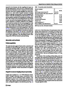
A 69-year-old woman with a history of coronary artery bypass surgery was hospitalized for the purpose of examining an aneurysm of the thoracic aorta. The patient reported back pain 2 weeks prior to admission. Transthoracic echocardiography with a 3.3 MHz probe from parasternal view showed dilated descending aorta (DAo) behind the left atrium. DAo was dilated to 4–5 cm and the left atrium was compressed slightly (Fig. 1a). Thickened inner wall of the aorta vessel was observed (Fig. 1b). Furthermore, CT image revealed the low density area which was not recognized by previous CT images. Therefore, she was diagnosed as non-communicating aortic dissection rather than mural thrombi, plaque, or artifacts. MRI examination was not performed. However, it was difficult to accurately identify whether it is plaque, non-communicating aortic dissection, or artifacts. We attempted paravertebral approach and were able to obtain excellent echo images with same echo probe (Fig. 1c).
This approach provided us information of non-communicating aortic dissection because color Doppler signal was not recognized onto a thickened part of the aorta (Fig. 1d). Furthermore, paravertebral approach showed vertically inverted parasternal window via DAo which is consistent with CT images (Fig. 1c, e, f). It is important to evaluate characteristic of aorta wall plaque and dissection [1–3]. Paravertebral approach is not usually used as a routine examination due to poor echo signal. However, because DAo becomes closer to the back skin in a patient with extremely dilated DAo, paravertebral approach enabled us to examine dilated characteristics of the aortic wall [4]. However, there can be a limitation in this approach because of inadequate dilatation of aorta for acute phase dissection. This approach may be a routine one for evaluation of dissection and aorta aneurysm.
* Tasuku Sato [email protected]‑u.ac.jp 1
Heart Center, Kyushu University Hospital, 3‑1‑1, Maidashi, Higashiku, Fukuoka 812‑8582, Japan
Department of Hematology, Oncology and Cardiovascular Medicine, Kyushu University Hospital, Fukuoka, Japan
2
13
Vol.:(0123456789)
Journal of Echocardiography
Fig. 1 Echo and CT imaging. Parasternal window was obtained from chest and paravertebral approach was obtained from the back. a, b Parasternal long and short axis view. There observed a dilated descending aorta and thickened inner wall of the aorta with suboptimal visibility. c Vertically inverted parasternal window via descending aorta. d Paravertebral approach can visualize thickened inner wall and confirm that there is no blood flow inside. e 3D CT imaging. (Red arrow: parasternal approach, Blue arrow: paravertebral approach). f Dilated descending aorta was approaching the wall of the back. DAo: Desc
Data Loading...