Incidental Diagnosis of an Arachnoid Granulation on Ga-68 DOTATATE PET/MRI
- PDF / 371,542 Bytes
- 3 Pages / 595.276 x 790.866 pts Page_size
- 23 Downloads / 225 Views
INTERESTING IMAGE
Incidental Diagnosis of an Arachnoid Granulation on Ga-68 DOTATATE PET/MRI Nghi C. Nguyen 1,2
&
Chan-Hong Moon 1 & Joseph M. Mettenburg 1
Received: 14 July 2020 / Revised: 12 August 2020 / Accepted: 17 August 2020 # Korean Society of Nuclear Medicine 2020
Keywords Arachnoid granulations . Pacchionian granulations . Meningiomas . Ga-68 DOTATATE PET . PET/MRI
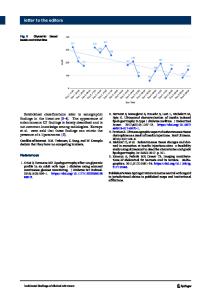
Arachnoid granulations (AGs), also known as Pacchionian granulations, are growths of the arachnoid membrane, allowing cerebrospinal fluid (CSF) to resorb from the subarachnoid space into the venous system. The growth of these AGs occasionally forms pits or remodeling of the skull base and calvarium. The prevalence of AGs on CT or MRI has been shown to vary dependent on the study populations and imaging techniques (e.g., MRI with or without MR angiography) as well as slice thickness of the imaging exams, given their occasionally small size. In one study, AGs were observed in 24% of patients undergoing contrast-enhanced CT and in 13% of contrast-enhanced MRI [1]. AGs and AG-associated pits tend to increase in size and number with age and exhibit similar imaging features to CSF on both CT and MRI. Characteristic MRI findings include isointense or even slightly hyperintense signal intensity relative to CSF on T1weighted imaging following Gd-based contrast agent administration (c, arrow), and bright signal intensity similar to CSF on T2 weighted sequences (d, arrow) [1, 2]. On CT, AG pits typically are observed as a well-circumscribed lesion with similar density to CSF that remodel the skull base or calvarium and may be perceived as an osteolytic lesion (f, arrow). AGs are also found within the dural venous sinuses causing a
* Nghi C. Nguyen [email protected] Chan-Hong Moon [email protected] Joseph M. Mettenburg [email protected] 1
Department of Radiology, University of Pittsburgh, Pittsburgh, PA, USA
2
Department of Radiology, UPMC Presbyterian, 200 Lothrop Street East Wing, Suite 200, Pittsburgh, PA 15213, USA
filling defect, commonly at insertion points of draining cortical veins. AGs have been shown to exhibit increased somatostatin receptor (SSTR), with all 12 tissue samples dissected from the superior sagittal sinus revealing equally strong SSTR subtypes 1 and 2 expressions [3]. The role of Ga-68 DOTATATE, a radioactive SSTR agonist, is well established for neuroendocrine tumors [4]. Moreover, it has added value for the diagnosis and clinical management of meningioma because of the high expression of SSTR subtype 2 in most cases [5, 6], and has been shown to have greater sensitivity compared with contrast-enhanced MRI [7–9]. Unlike AGs, meningiomas are typically duralbased mass lesions, isointense to gray matter on both T1and T2-weighted MRI and demonstrate brisk enhancement after Gd-based contrast agent administration. Meningiomas can occasionally be intra-osseous or even extra-cranial. It is postulated that meningiomas arise from arachnoid cap cells and commonly occur where these cells are most numerous, such as in areas of AGs [
Data Loading...